Copyright
©The Author(s) 2020.
World J Gastroenterol. Oct 28, 2020; 26(40): 6295-6303
Published online Oct 28, 2020. doi: 10.3748/wjg.v26.i40.6295
Published online Oct 28, 2020. doi: 10.3748/wjg.v26.i40.6295
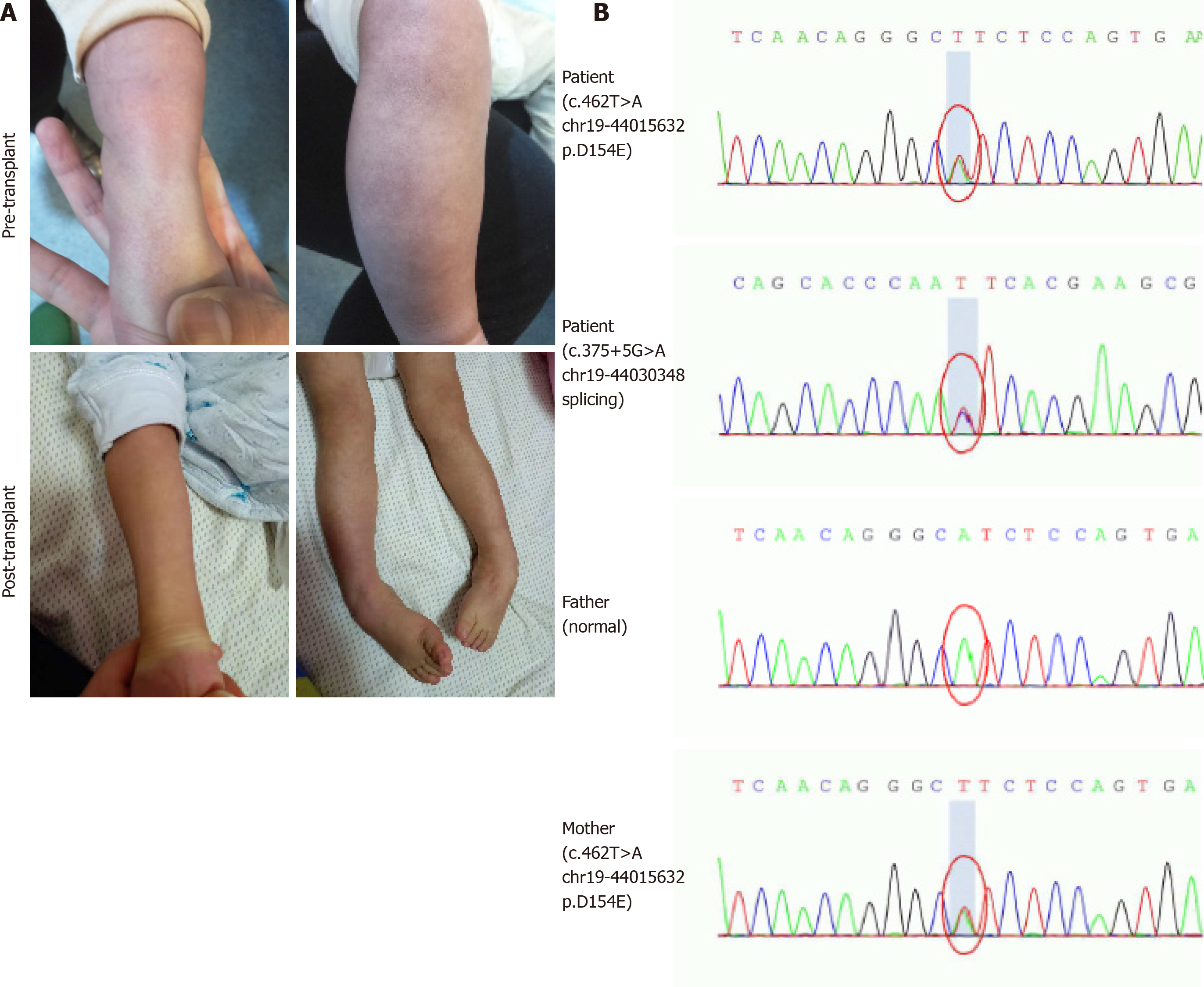
Figure 1 Clinical symptoms and gene sequencing analysis.
A: The pre- and post-transplant scattered petechiae in the extremities and orthostatic acrocyanosis; B: Sequence electropherogram of the compound heterozygous mutation (c.375+5G>A and c.462T>A) in patient, heterozygous mutation in mother, and no mutation in father.
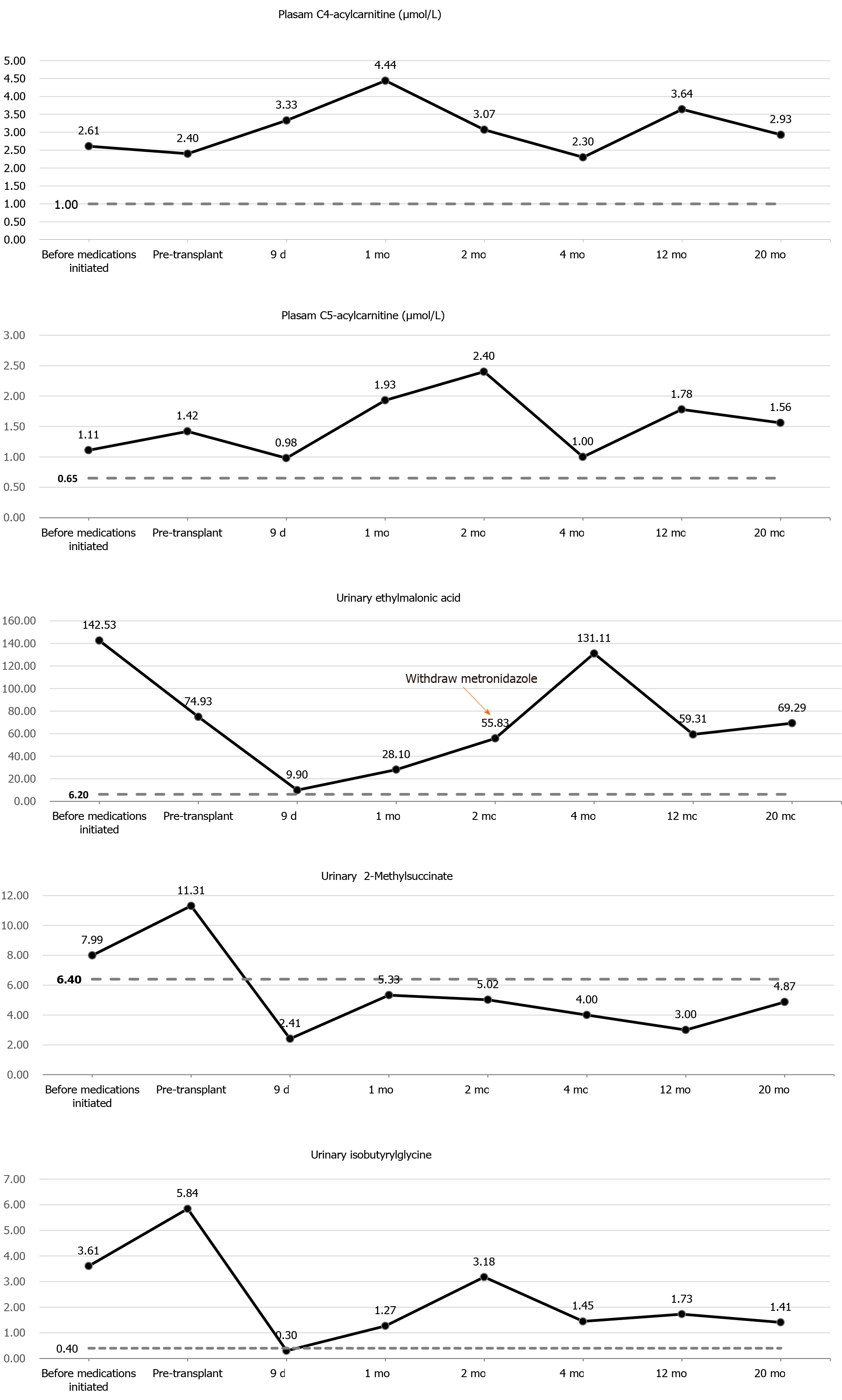
Figure 2 Changes of pre- and post-transplant urine organic acid profile and plasma acylcarnitine profile in the patient with ethylmalonic encephalopathy.
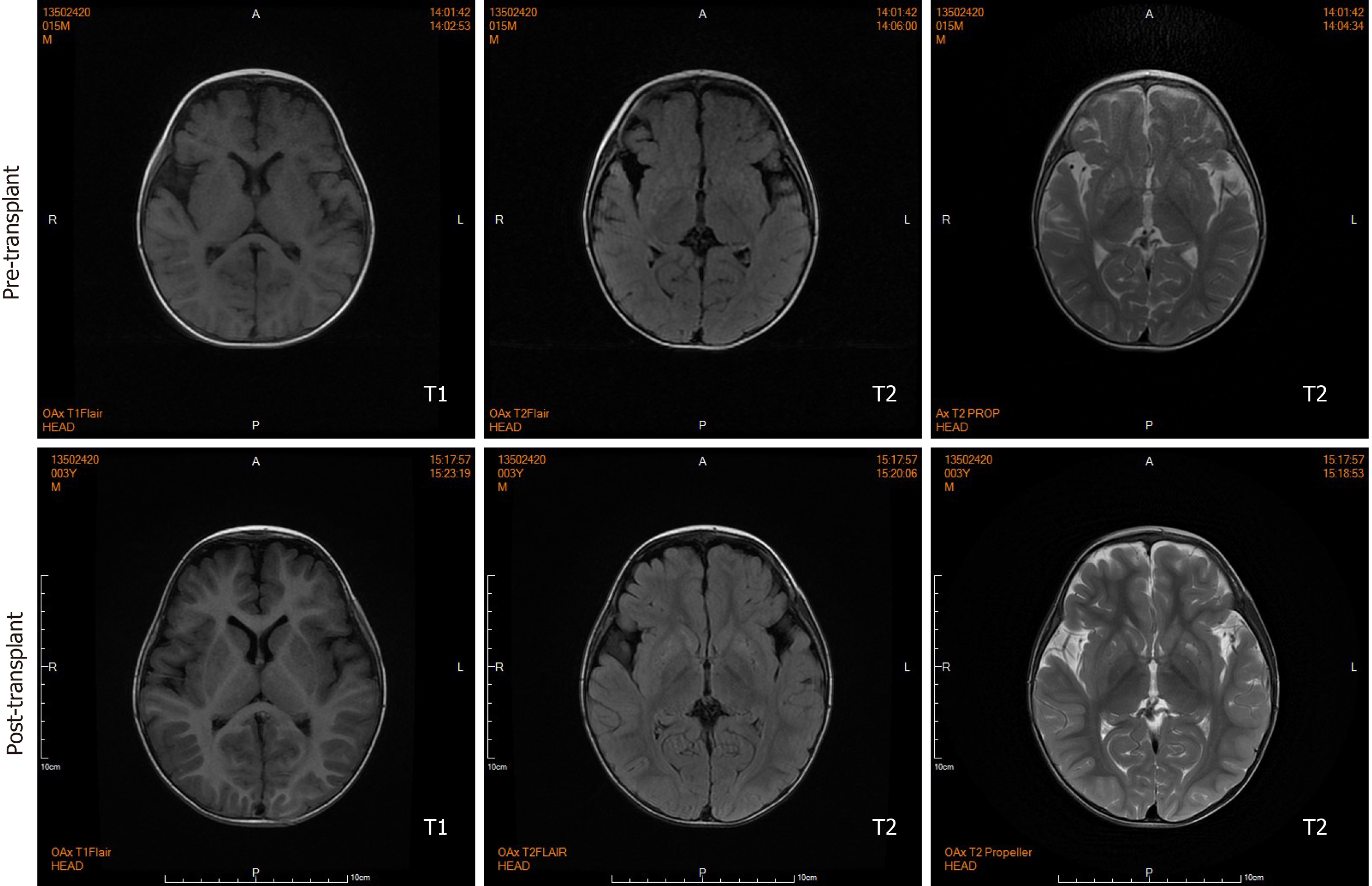
Figure 3 Pre- and post-transplant axial sections of brain magnetic resonance imaging scan.
Before liver transplantation, brain magnetic resonance imaging shows fronto-temporal atrophy with multiple bilateral symmetrical abnormal low and high signal intensity involving caudate nucleus and lentiform nucleus in T1- and T2-weighted sections, respectively. Twenty months after transplantation, brain magnetic resonance imaging shows lesions remained but ameliorated.
- Citation: Zhou GP, Qu W, Zhu ZJ, Sun LY, Wei L, Zeng ZG, Liu Y. Compromised therapeutic value of pediatric liver transplantation in ethylmalonic encephalopathy: A case report. World J Gastroenterol 2020; 26(40): 6295-6303
- URL: https://www.wjgnet.com/1007-9327/full/v26/i40/6295.htm
- DOI: https://dx.doi.org/10.3748/wjg.v26.i40.6295


