Copyright
©The Author(s) 2020.
World J Gastroenterol. Jan 21, 2020; 26(3): 353-365
Published online Jan 21, 2020. doi: 10.3748/wjg.v26.i3.353
Published online Jan 21, 2020. doi: 10.3748/wjg.v26.i3.353
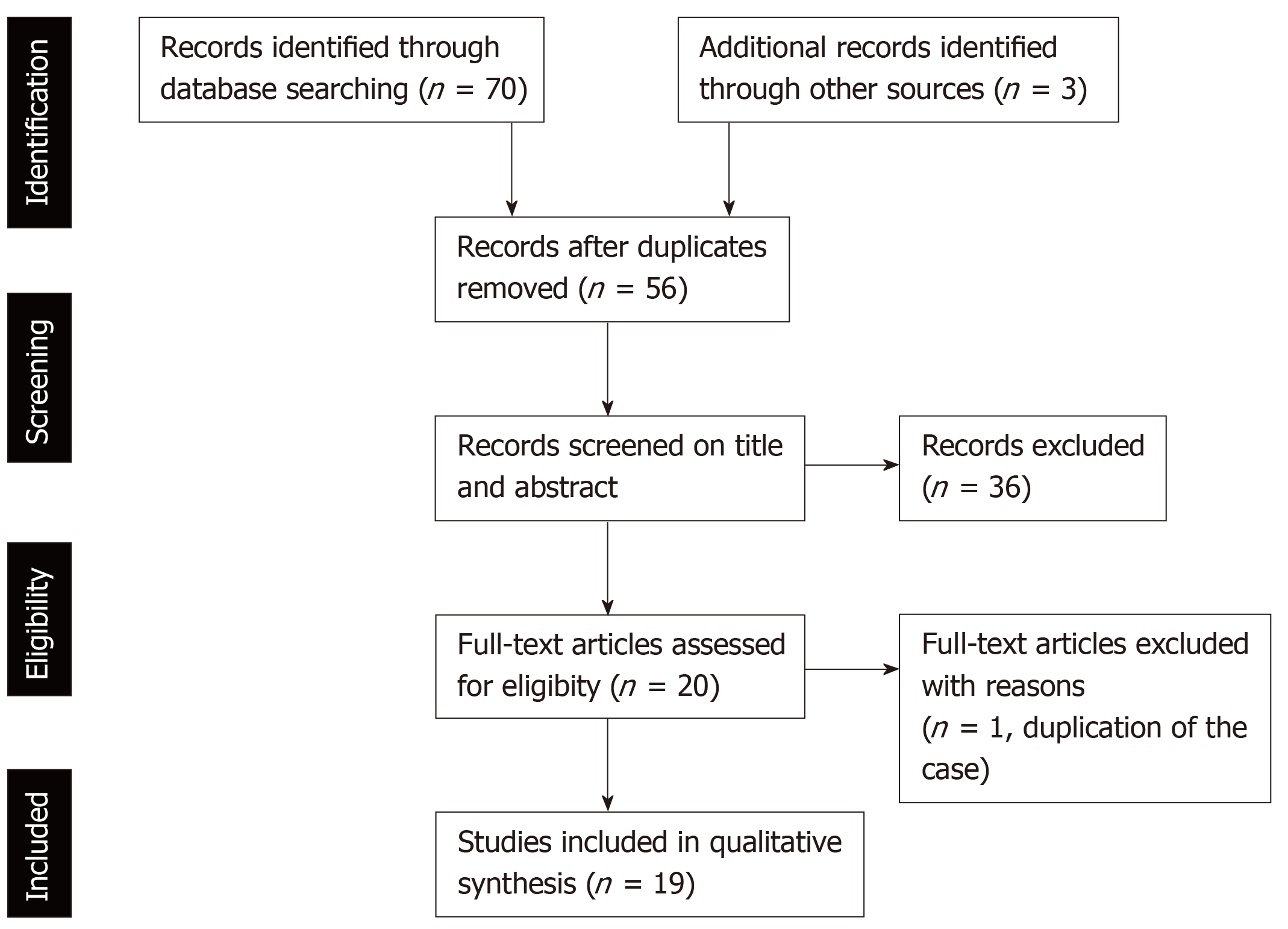
Figure 1 PRISMA 2009 Flow diagram describing the selection of the studies reporting programmed cell death-1 inhibitor-related cholangitis for our review.
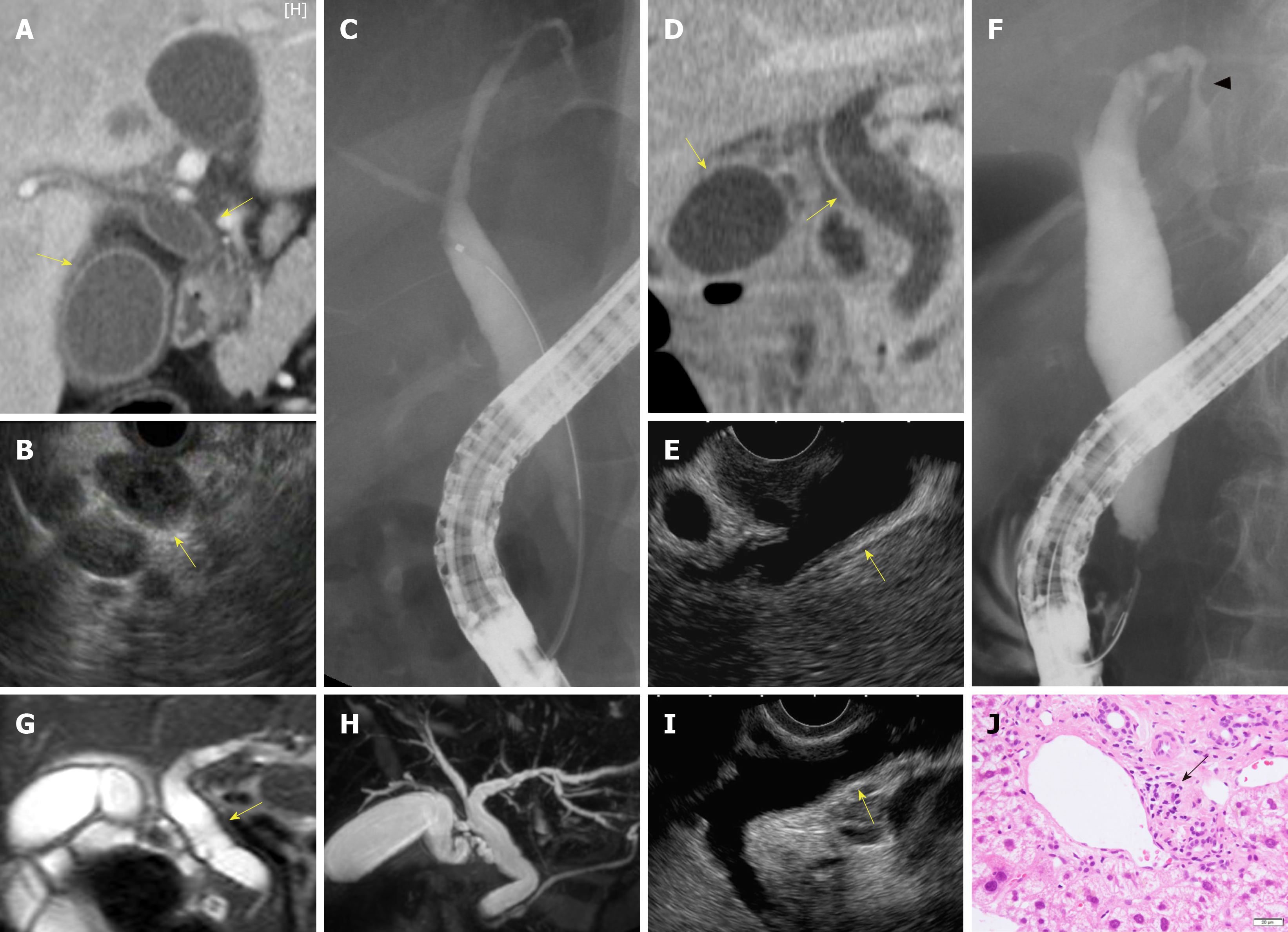
Figure 2 Our cases with sclerosing cholangitis caused by nivolumab (Case 29, A–C; Case 30, D–F; Case 31, G–J).
A, D: Computed tomography; G, H: Magnetic resonance imaging; B, E, I: Endoscopic ultrasonography. Revealed diffuse hypertrophy of biliary tract (yellow allows) in all cases. C, F: Endoscopic retrograde cholangiopancreatography revealed biliary stenosis of the intrahepatic bile duct (arrow-head) in Case 30. J: In Case 31, liver biopsy showed lymphocyte infiltration in Glisson’s capsule (black arrow).
- Citation: Onoyama T, Takeda Y, Yamashita T, Hamamoto W, Sakamoto Y, Koda H, Kawata S, Matsumoto K, Isomoto H. Programmed cell death-1 inhibitor-related sclerosing cholangitis: A systematic review. World J Gastroenterol 2020; 26(3): 353-365
- URL: https://www.wjgnet.com/1007-9327/full/v26/i3/353.htm
- DOI: https://dx.doi.org/10.3748/wjg.v26.i3.353


