Copyright
©The Author(s) 2019.
World J Gastroenterol. Aug 7, 2019; 25(29): 3941-3955
Published online Aug 7, 2019. doi: 10.3748/wjg.v25.i29.3941
Published online Aug 7, 2019. doi: 10.3748/wjg.v25.i29.3941
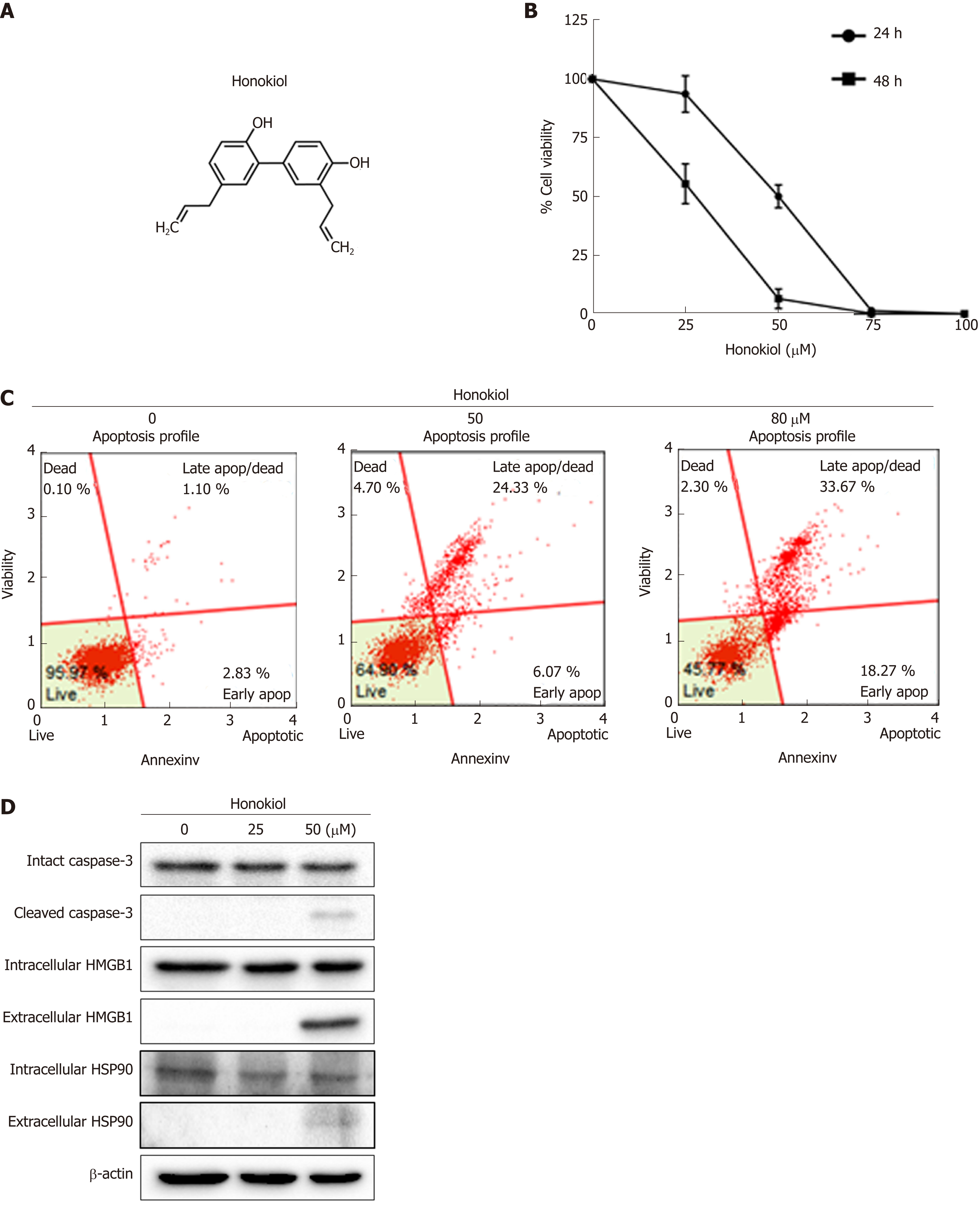
Figure 1 Honokiol induced cholangiocarcinoma cell apoptosis and cause of Damage- associated molecular pattern secretion.
A: Chemical structure of honokiol; B: KKU-213L5 cells were treated with various concentrations of honokiol. After 24 and 48 h, cell viability was accessed using MTT assay. Negative control was cells treated with dimethyl sulfoxide and all groups were normalised with the control group. Results were presented as mean ± SD of three independent experiments; C: Treated KKU-213L5 cells were analyzed using Muse™ Cell Analyser with annexin V/PI staining. Annexin V versus propidium iodide from the gated cells revealed cell populations as live, early apoptotic, late apoptotic/dead and dead; D: Protein samples and conditioned medium were collected and analysed using Western blot. HMGB1: High mobility group box1; HSP: Heat shock protein.
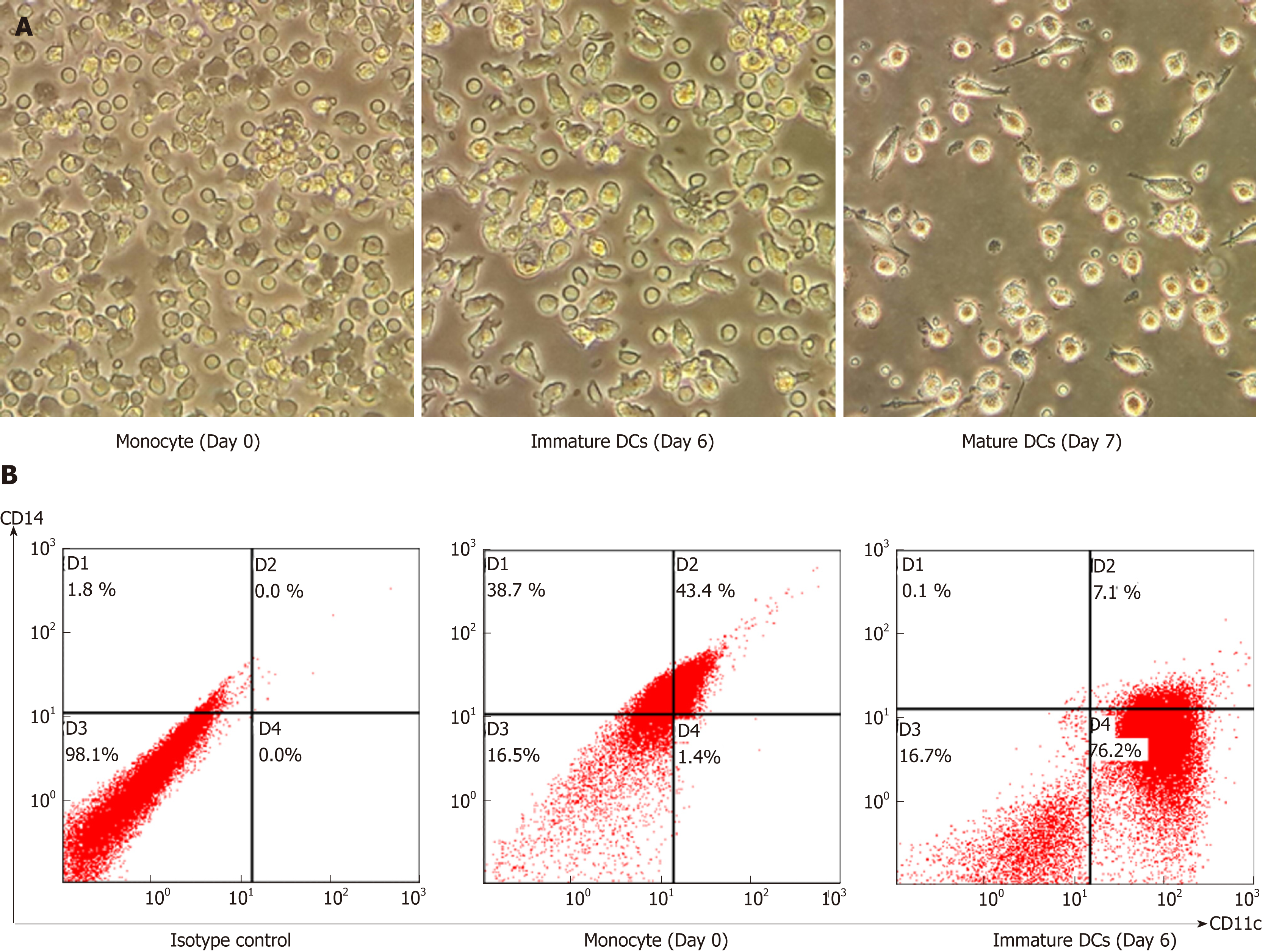
Figure 2 Morphology and immunophenotype of human monocyte-derived dendritic cells.
Human monocytes were cultured in RPMI1640 medium supplemented with granulocyte-macrophage colony-stimulating factor and interleukin-4 for 6 d. Immature dendritic cells were then stimulated to become mature by adding tumor necrosis factor-α and interferon-γ for 1 d. A: Cell morphology of monocytes. Immature and mature dendritic cells were observed under inverted microscopy at 40×; B: Expressions of CD14 and CD11c human monocytes and mature DCs were presented as percentage of gated cells using flow cytometry CXP software. DCs: Dendritic cells.
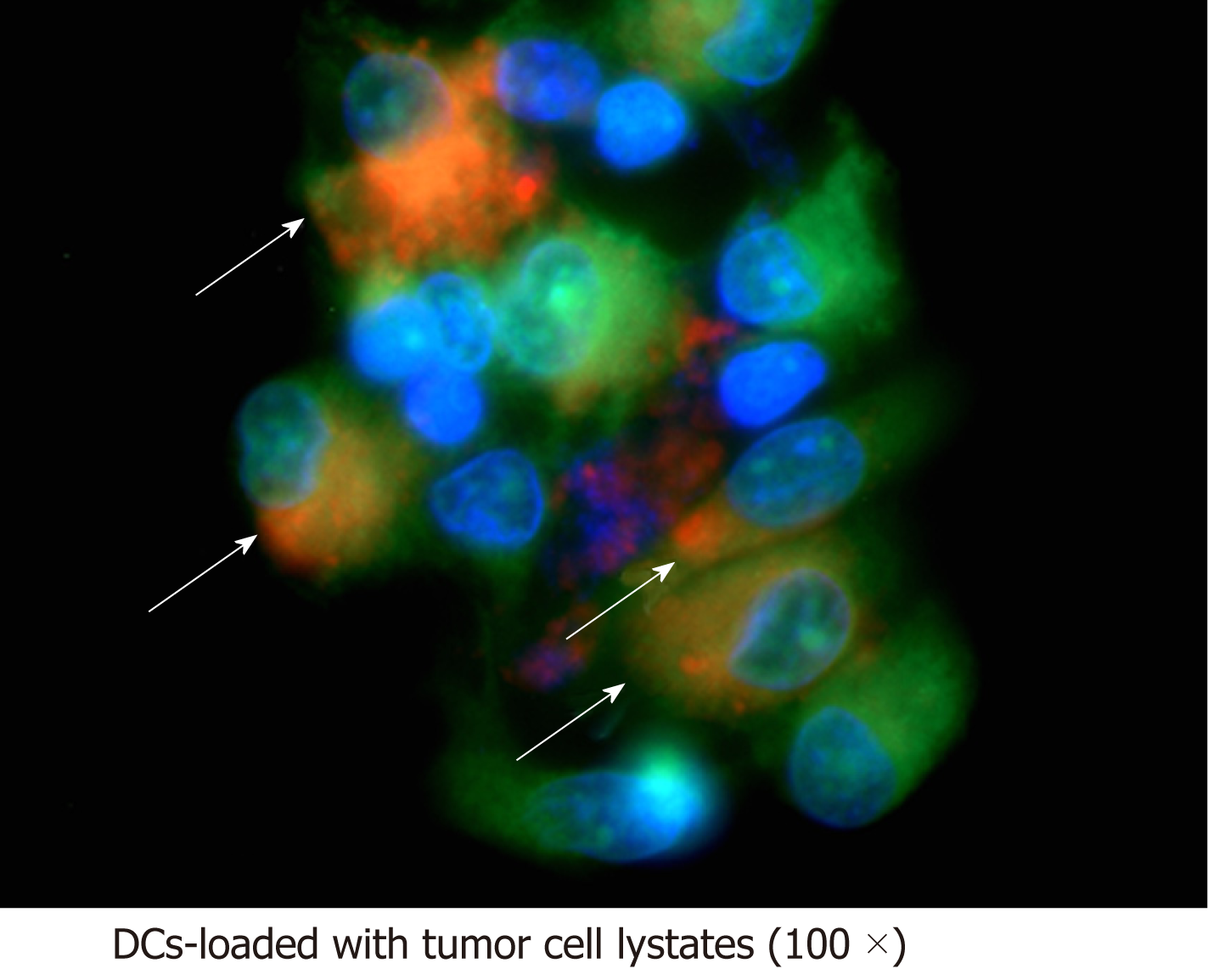
Figure 3 Investigation of tumour antigen on dendritic cells by fluorescence microscopy.
Dendritic cells were stained with CellTracker™ Green CMFDA before loading with honokiol-derived tumour cell lysates pre-stained with CellTracker™ Red CMPTX. Antigen load was verified by visualisation of tumour antigen (red) within dendritic cells (green). Arrowheads indicate co-expression of green and red fluorescence. DCs: Dendritic cells.
Figure 4 Honokiol-derived tumour cell lysates pulsed dendritic cells significantly stimulated T lymphocyte proliferation.
KKU-213L5 cells were pretreated with honokiol or left untreated before they were lysed and loaded into dendritic cells (DCs). T lymphocytes were stimulated with different types of DCs (unpulsed, pulsed with tumour cell lysates and pulsed with honokiol-derived tumour cell lysates) at 1:10 ratios for 5 d. Lymphocyte number was reflected by direct counting (A) and relative lymphocyte proliferation (B). T lymphocytes cultured alone were set as control and all experiments were performed in triplicate. Results are shown as mean ± SD, A P value < 0.05 was considered significant; aP < 0.05, bP < 0.01 and cP < 0.001. DCs: Dendritic cells; HOK: Honokiol; TCL: Tumour cell lysate.
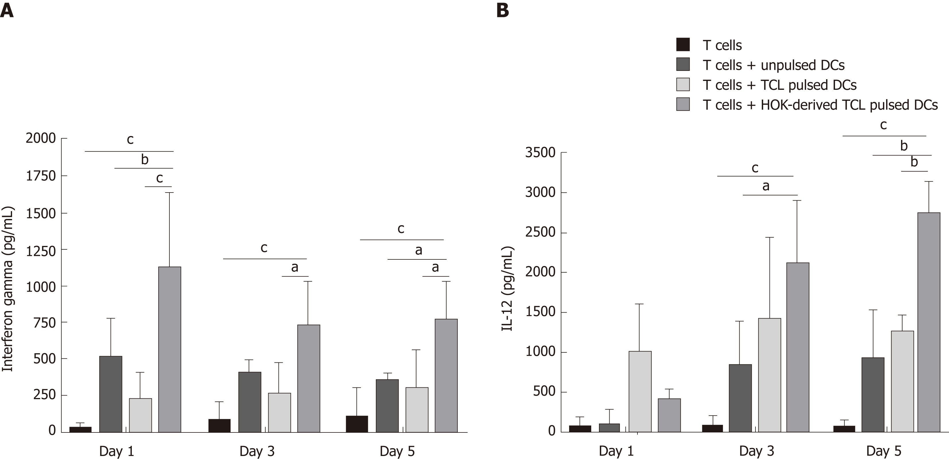
Figure 5 Type I cytokines interferon-γ and interleukin-12 production.
Autologous T lymphocytes were cocultured either with unloaded or loaded tumour cell lysates or tumour cell lysates derived from honokiol-treated CCA cells for 1-5 days before measurement of cytokines. Levels of interferon-γ (A) and interleukin-12 (B) in conditioned medium were analysed by ELISA. Results are shown as mean ± SD of three independent experiments. A P value < 0.05 was considered significant; aP < 0.05, bP < 0.01 and cP < 0.001. DCs: Dendritic cells; HOK: Honokiol; TCL: Tumour cell lysate.
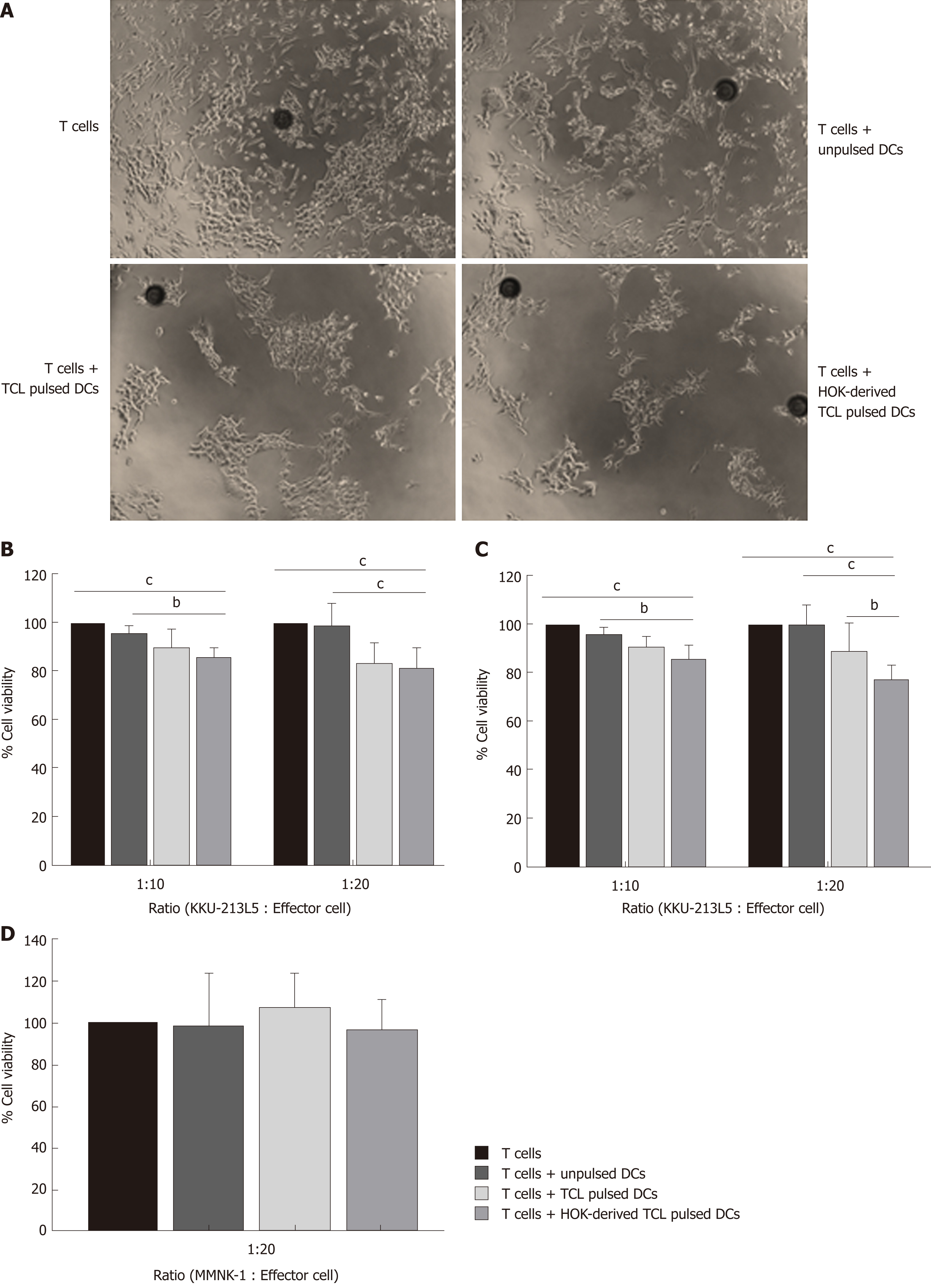
Figure 6 Dendritic cells pulsed with tumour cell lysates derived from honokiol-treated cholangiocarcinoma cells significantly enhanced T lymphocyte killing of cholangiocarcinoma cells.
Differently primed effector T lymphocytes were cocultured with target KKU-213L5 cell line at ratios of 1:10 and 1:20. Killing capacity was evaluated using the MTT assay. A: Remaining target cells after coculture with effector cells for 48 h at 1:20 ratio photographed at 100×. Cytotoxic effect of T lymphocytes on KKU-213L5 is shown as % viability of target cells at (B) 24 h, (C) 48 h and for MMNK1 (D) at 48 h. Results are presented as mean ± SD of three independent experiments. A P value < 0.05 was considered significant; aP < 0.05, bP < 0.01 and cP < 0.001. DCs: Dendritic cells; HOK: Honokiol; TCL: Tumour cell lysate.
- Citation: Jiraviriyakul A, Songjang W, Kaewthet P, Tanawatkitichai P, Bayan P, Pongcharoen S. Honokiol-enhanced cytotoxic T lymphocyte activity against cholangiocarcinoma cells mediated by dendritic cells pulsed with damage-associated molecular patterns. World J Gastroenterol 2019; 25(29): 3941-3955
- URL: https://www.wjgnet.com/1007-9327/full/v25/i29/3941.htm
- DOI: https://dx.doi.org/10.3748/wjg.v25.i29.3941


