Copyright
©The Author(s) 2018.
World J Gastroenterol. Jul 21, 2018; 24(27): 2921-2930
Published online Jul 21, 2018. doi: 10.3748/wjg.v24.i27.2921
Published online Jul 21, 2018. doi: 10.3748/wjg.v24.i27.2921
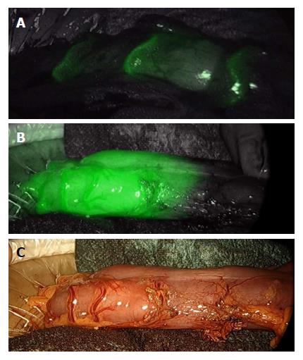
Figure 1 Colon perfusion before anastomosis during left colectomy.
A few seconds after the i.v. injection of 0.3 mg/kg indocyanine green, bowel arteries clearly appear (A); thereafter, the bowel perfusion cut-off area becomes evident (B and C).
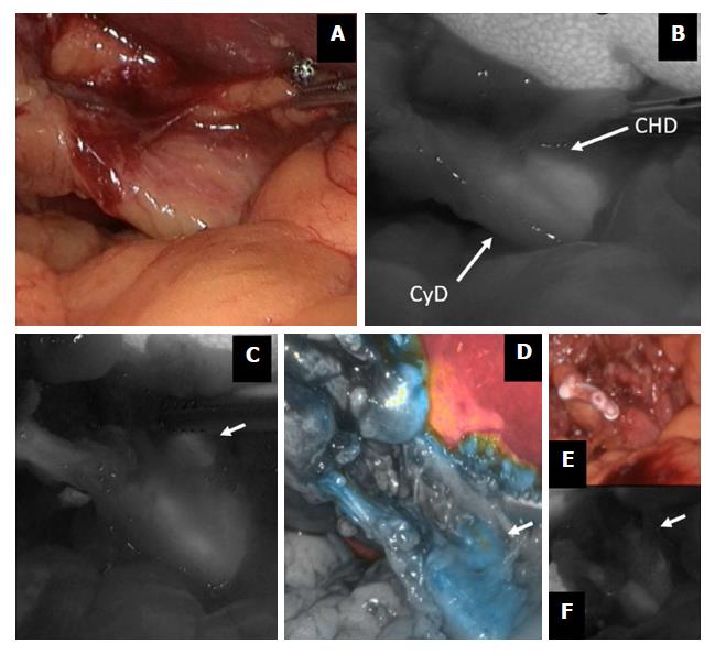
Figure 2 Indocyanine green-enhanced biliary anatomy.
During a difficult cholecystectomy for acute cholecystitis (A), the confluence between the cystic duct (CyD) and the common hepatic duct (CHD) is shown by fluorescence imaging (B); common hepatic duct (arrow) is further visualized before (C and D) and after (E and F) cystic duct division. ICG: Indocyanine green.
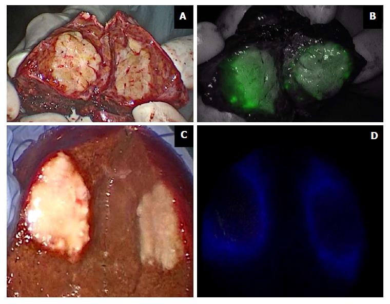
Figure 3 Indocyanine green in liver surgery.
Primary liver tumors show intense and complete staining because their hepatocytes take up ICG but do not secrete it (A and B); liver metastases show a ring appearance because their cells do not take up ICG but hepatocytes surrounding the nodule are compressed (C and D). ICG: Indocyanine green.
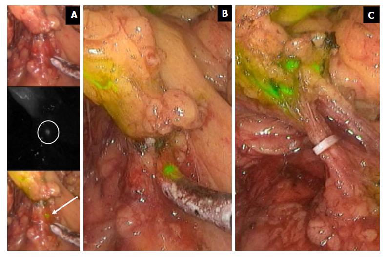
Figure 4 Indocyanine green fluorescence imaging in extended right hemicolectomy.
The figure displays the right branches of middle colic vessel division during extended right hemicolectomy for transverse colon cancer. ICG injected in the tumor site spreads in nodes at the very proximal root of the artery. ICG fluorescence imaging allows a radical lymphadenectomy, including very small nodes (A and B). Only when all the stained nodes are removed may the nodal dissection be considered radical (C). ICG: Indocyanine green.
- Citation: Baiocchi GL, Diana M, Boni L. Indocyanine green-based fluorescence imaging in visceral and hepatobiliary and pancreatic surgery: State of the art and future directions. World J Gastroenterol 2018; 24(27): 2921-2930
- URL: https://www.wjgnet.com/1007-9327/full/v24/i27/2921.htm
- DOI: https://dx.doi.org/10.3748/wjg.v24.i27.2921


