Copyright
©The Author(s) 2017.
World J Gastroenterol. Feb 7, 2017; 23(5): 906-918
Published online Feb 7, 2017. doi: 10.3748/wjg.v23.i5.906
Published online Feb 7, 2017. doi: 10.3748/wjg.v23.i5.906
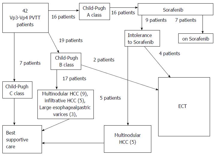
Figure 1 Selection of eligible patients to electrochemotherapy among a consecutive series of 42 patients with VP3-VP4 portal vein tumor thrombosis.
ECT: Electrochemotherapy; PVTT: Portal vein tumor thrombosis; HCC: Hepatocellular carcinoma.
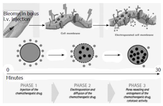
Figure 2 Illustrative sketch of the mechanism with which electrochemotherapy operates: After insertion of the electrodes in the tissue, a bleomycin bolus is administered intravenously.
Eight minutes after injection, bleomycin diffuses in the tumor tissue. At this point the electric pulses are started in order to alter permeability of cells’ membranes. The electroporation process greatly increases the bleomycin intracellular entrance. Therefore by self repair of the membranes, pores reseal and the bleomycin is entrapped in the cells.
Figure 3 Insertion of six electrodes for electrochemotherapy treatment of right portal vein.
A: Illustrative sketch showing schematically the position of electrodes around the external margin of the Tumor thrombosis; B: Up to six electrode-needles are inserted percutaneously; C: The position of electrodes can be monitored with US during the procedure.
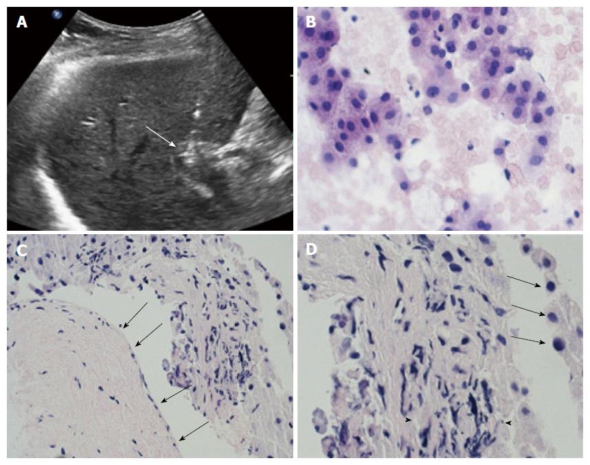
Figure 4 All patients underwent pre- and post-treatment biopsy of the portal vein tumor thrombus.
A: ultrasound scan demonstrate the correct positioning of the needle tip (arrow) in the thrombus; B: Intraoperative pre-treatment biopsy of the thrombus was adequate and showed viable cells from hepatocellular carcinoma in 5/6 cases; C: High magnification of biopsy specimen showed severe involutive changes of tumor cells with cellular apoptosis (arrows) and areas of necrosis (arrowheads) in all six cases. Low magnification in the same specimen, beside the altered tumor thrombus, showed absence of damage from the procedure to portal vein wall (arrows); D: Portal endothelium shows normal appearance with regular wall layers.
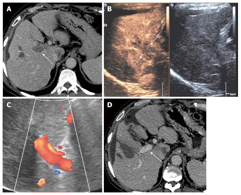
Figure 5 M.
D.S. 64 years hepatitis C virus related cirrhosis. A: On september 2014 computed tomography (CT) showed complete malignant thrombosis of right portal vein (arrows); B: Pretreatment contrast enhanced ultrasound showed diffuse enhancement of the thrombus (arrows) consistent with malignant thrombosis; The patient underwent electrochemotherapy with insertion of 6 electrode-needles and i.v. Bleomicin bolus; C: Three months monthly color-Doppler ultrasound control showed complete patency of the right portal vein; D: Persistent patency of right portal vein (arrows) and absence of local recurrence was confirmed at 3, 9, 15 mo follow-up CT control.
- Citation: Tarantino L, Busto G, Nasto A, Fristachi R, Cacace L, Talamo M, Accardo C, Bortone S, Gallo P, Tarantino P, Nasto RA, Di Minno MND, Ambrosino P. Percutaneous electrochemotherapy in the treatment of portal vein tumor thrombosis at hepatic hilum in patients with hepatocellular carcinoma in cirrhosis: A feasibility study. World J Gastroenterol 2017; 23(5): 906-918
- URL: https://www.wjgnet.com/1007-9327/full/v23/i5/906.htm
- DOI: https://dx.doi.org/10.3748/wjg.v23.i5.906


