Copyright
©The Author(s) 2016.
World J Gastroenterol. Apr 14, 2016; 22(14): 3879-3884
Published online Apr 14, 2016. doi: 10.3748/wjg.v22.i14.3879
Published online Apr 14, 2016. doi: 10.3748/wjg.v22.i14.3879
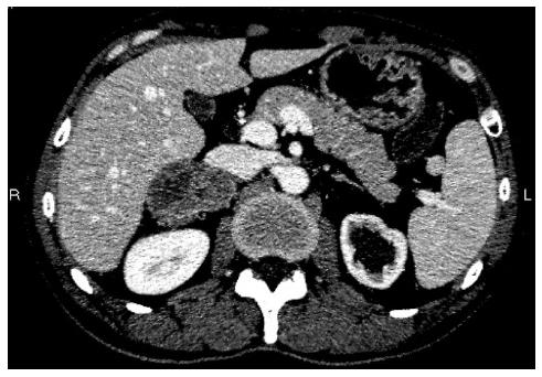
Figure 1 Abdominal enhanced computed tomography revealed a low density mass in the right adrenal gland.
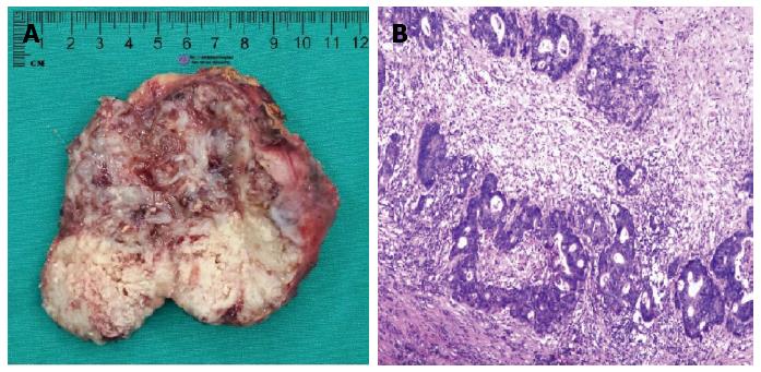
Figure 2 Adrenal tumor.
A: Macroscopic findings of the adrenal lesion showed a solid tumor with necrosis; B: Microscopic findings of the adrenal tumor showed moderately differentiated adenocarcinoma that resembles the primary sigmoid lesion that had been resected. This indicated that the metastasis was from the sigmoid carcinoma.
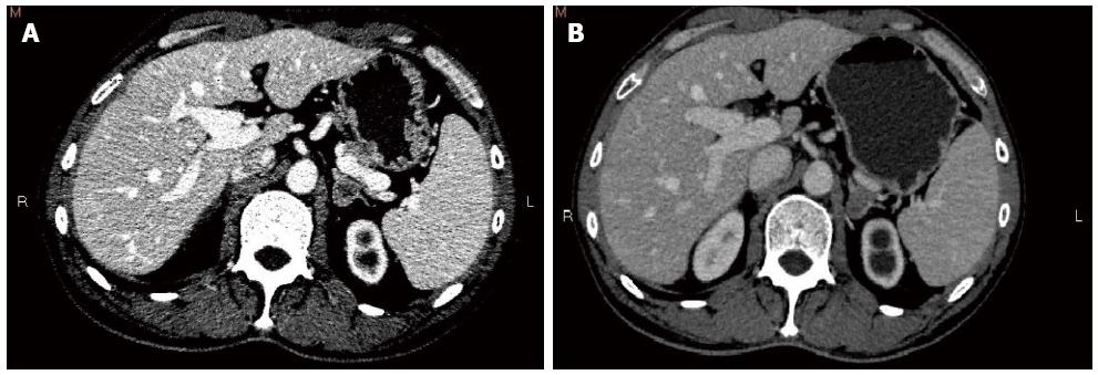
Figure 3 Computed tomography image.
A: Computed tomography (CT) of the abdomen shows a left adrenal mass; B: CT evaluation showed response of the adrenal metastases after chemotherapy.
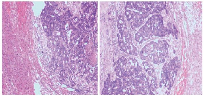
Figure 4 Pathological findings of the left adrenal tumor showed moderately differentiated adenocarcinoma, compatible with metastasis from the rectal carcinoma.
- Citation: Liu YY, Chen ZH, Zhai ET, Yang J, Xu JB, Cai SR, Song W. Case of metachronous bilateral isolated adrenal metastasis from colorectal adenocarcinoma and review of the literature. World J Gastroenterol 2016; 22(14): 3879-3884
- URL: https://www.wjgnet.com/1007-9327/full/v22/i14/3879.htm
- DOI: https://dx.doi.org/10.3748/wjg.v22.i14.3879


