
















©The Author(s) 2016.
World J Gastroenterol. Apr 14, 2016; 22(14): 3879-3884
Published online Apr 14, 2016. doi: 10.3748/wjg.v22.i14.3879
Published online Apr 14, 2016. doi: 10.3748/wjg.v22.i14.3879
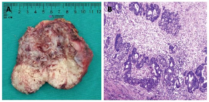
Figure 2 Adrenal tumor.
A: Macroscopic findings of the adrenal lesion showed a solid tumor with necrosis; B: Microscopic findings of the adrenal tumor showed moderately differentiated adenocarcinoma that resembles the primary sigmoid lesion that had been resected. This indicated that the metastasis was from the sigmoid carcinoma.
- Citation: Liu YY, Chen ZH, Zhai ET, Yang J, Xu JB, Cai SR, Song W. Case of metachronous bilateral isolated adrenal metastasis from colorectal adenocarcinoma and review of the literature. World J Gastroenterol 2016; 22(14): 3879-3884
- URL: https://www.wjgnet.com/1007-9327/full/v22/i14/3879.htm
- DOI: https://dx.doi.org/10.3748/wjg.v22.i14.3879