Copyright
©The Author(s) 2015.
World J Gastroenterol. Feb 21, 2015; 21(7): 2210-2213
Published online Feb 21, 2015. doi: 10.3748/wjg.v21.i7.2210
Published online Feb 21, 2015. doi: 10.3748/wjg.v21.i7.2210
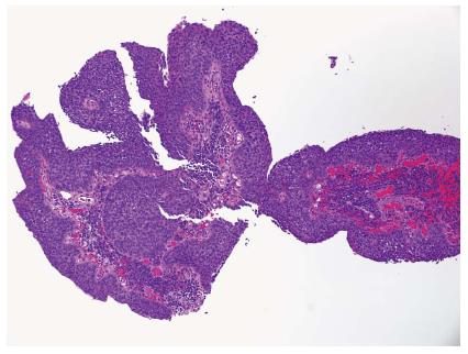
Figure 1 Biopsy of anal mass showing a squamous cell carcinoma with prominent papillary features (HE, × 100).
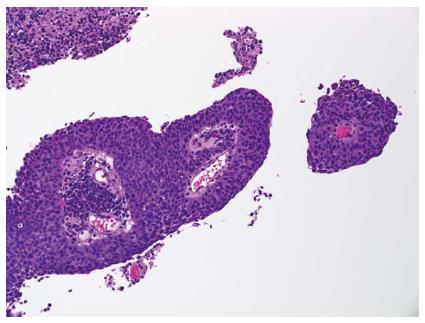
Figure 2 Higher power view of the papillary structures and associated anal intraepithelial neoplasia 3 (carcinoma-situ) (HE, × 200).
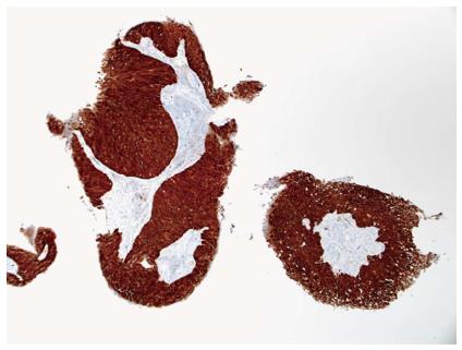
Figure 3 p-16 immunohistochemical stain showing strong and diffuse positive reaction of the tumor cells (× 100).
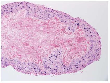
Figure 4 In-situ hybridization for high risk types of human papillomavirus showing a strong and diffuse reaction of the epithelial tumor cells (× 200).
- Citation: Leon ME, Shamekh R, Coppola D. Human papillomavirus-related squamous cell carcinoma of the anal canal with papillary features. World J Gastroenterol 2015; 21(7): 2210-2213
- URL: https://www.wjgnet.com/1007-9327/full/v21/i7/2210.htm
- DOI: https://dx.doi.org/10.3748/wjg.v21.i7.2210


