Copyright
©2013 Baishideng Publishing Group Co.
World J Gastroenterol. Jan 14, 2013; 19(2): 307-310
Published online Jan 14, 2013. doi: 10.3748/wjg.v19.i2.307
Published online Jan 14, 2013. doi: 10.3748/wjg.v19.i2.307
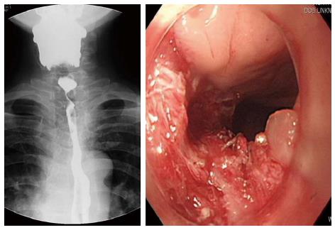
Figure 1 Esophagogram and endoscopic findings before patch esophagoplasty.
The native esophagus was visualized with the esophagogram, but the colon conduit from bypass surgery was not visible. After insertion of an Endoscopic Varix Ligation tube, a stenotic lesion of the esophago-colonic anastomosis site was opened. Focal luminal stenosis by fibrosis was evident. Endoscopic dilatation was repeated using electrocauterization.
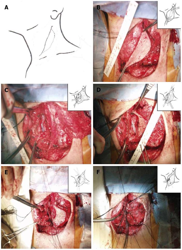
Figure 2 Photograph of the musculocutaneous sternocleidomastoid flap and esophagotomy during the operation.
A: A skin incision was designed for an elliptical skin island based on the sternocleidomastoid muscle; B: A skin island (6 cm × 4 cm) on the sternocleidomastoid (SCM) muscle was prepared; C, D: Esophagotomy at the level of the cricoid cartilage was extended upward to pharynx and downward; E: The SCM muscle was severed near the clavicle and rotated. The skin flap was fixated to esophagus with interrupted silk sutures; F: The esophagoplasty site was covered with SCM muscle via multiple fixation sutures.
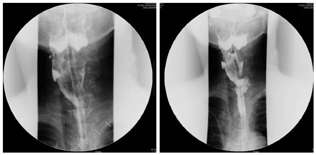
Figure 3 Esophagogram after sternocleidomastoid myocutaneous patch esphagoplasty.
The stenotic lesion at the esophago-colonic anastomosis site was widened successfully and the dye passed through the colon conduit without resistance.
- Citation: Sa YJ, Kim YD, Kim CK, Park JK, Moon SW. Recurrent cervical esophageal stenosis after colon conduit failure: Use of myocutaneous flap. World J Gastroenterol 2013; 19(2): 307-310
- URL: https://www.wjgnet.com/1007-9327/full/v19/i2/307.htm
- DOI: https://dx.doi.org/10.3748/wjg.v19.i2.307


