Copyright
©2013 Baishideng Publishing Group Co.
World J Gastroenterol. Jan 14, 2013; 19(2): 307-310
Published online Jan 14, 2013. doi: 10.3748/wjg.v19.i2.307
Published online Jan 14, 2013. doi: 10.3748/wjg.v19.i2.307
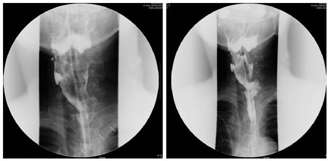
Figure 3 Esophagogram after sternocleidomastoid myocutaneous patch esphagoplasty.
The stenotic lesion at the esophago-colonic anastomosis site was widened successfully and the dye passed through the colon conduit without resistance.
- Citation: Sa YJ, Kim YD, Kim CK, Park JK, Moon SW. Recurrent cervical esophageal stenosis after colon conduit failure: Use of myocutaneous flap. World J Gastroenterol 2013; 19(2): 307-310
- URL: https://www.wjgnet.com/1007-9327/full/v19/i2/307.htm
- DOI: https://dx.doi.org/10.3748/wjg.v19.i2.307


