Copyright
©2012 Baishideng Publishing Group Co.
World J Gastroenterol. Nov 14, 2012; 18(42): 6168-6171
Published online Nov 14, 2012. doi: 10.3748/wjg.v18.i42.6168
Published online Nov 14, 2012. doi: 10.3748/wjg.v18.i42.6168
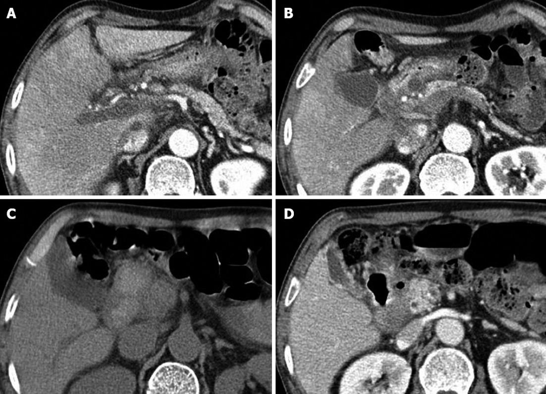
Figure 1 Abdominal computed tomography images.
A: A filling defect and intra-luminal hypoattenuation in the portal vein consistent with acute portal vein thrombosis; B: A 12 mm × 10 mm cystic lesion suggestive of a pseudocyst and extensive inflammatory fluid collections around the pancreatic head; C: A walled-in fluid collection with high attenuation, indicative findings of a hemorrhagic pseudocyst around the pancreatic head and compressed duodenum; D: Reduced hemorrhagic fluid collections and an air-filled cyst, indicative of a fistula between the duodenum and the pseudocyst.
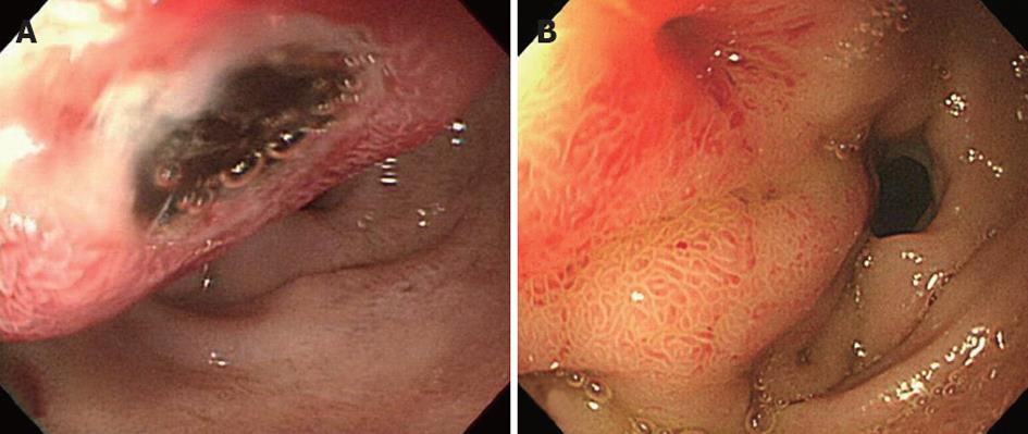
Figure 2 Upper gastrointestinal endoscopy.
A: Emergency endoscopy shows a 2 cm submucosal tumor-like bulging mass with central ulceration; B: A follow-up endoscopy image showing a decreased mass size and fistula orifice.
- Citation: Park WS, Kim HI, Jeon BJ, Kim SH, Lee SO. Should anticoagulants be administered for portal vein thrombosis associated with acute pancreatitis? World J Gastroenterol 2012; 18(42): 6168-6171
- URL: https://www.wjgnet.com/1007-9327/full/v18/i42/6168.htm
- DOI: https://dx.doi.org/10.3748/wjg.v18.i42.6168


