Copyright
©2009 The WJG Press and Baishideng.
World J Gastroenterol. Feb 21, 2009; 15(7): 804-816
Published online Feb 21, 2009. doi: 10.3748/wjg.15.804
Published online Feb 21, 2009. doi: 10.3748/wjg.15.804
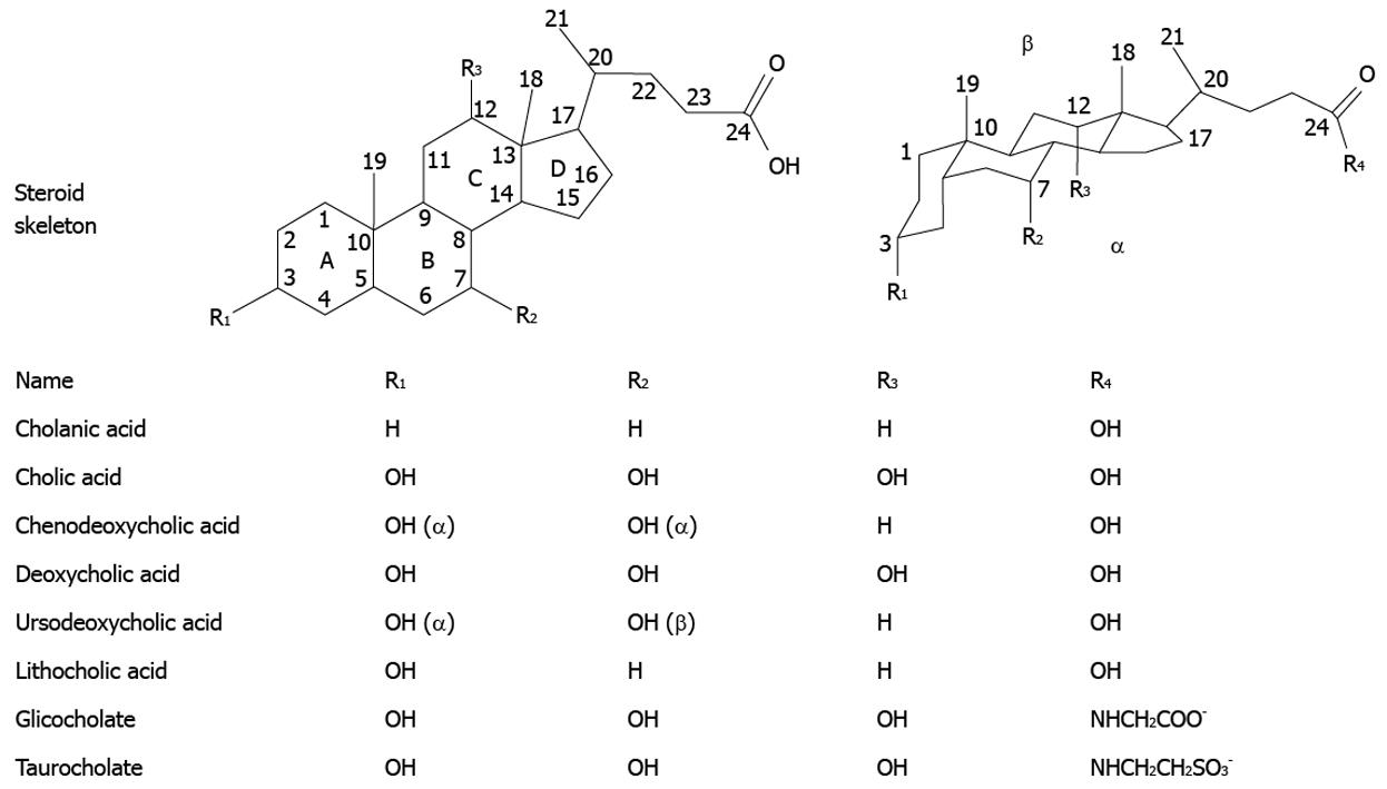
Figure 1 Structures of the most abundant bile acids in humans, and their glycine and taurine conjugates.
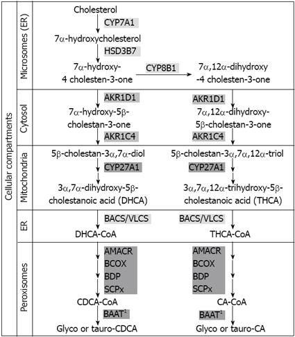
Figure 3 Schematic representation of bile acid synthesis by the classical neutral pathway.
AKR1C4: 3α-hydroxysteroid dehydrogenase; AKR1D1: Δ4–3-oxosteroid-5β-reductase; AMACR: Alpha methylacyl-CoA racemase; BAAT: Bile acid; CoA: Amino acid N-acyltransferase (1A minor cytosolic fraction does also exist); BACS: Bile acid CoA synthetase; BCOX: Branched-chain acyl CoA oxidase; BDP: D-bifunctional protein hydratase; CYP27A1: Sterol 27-hydroxylase; CYP7A1: Cholesterol 7α-hydroxylase; CYP8B1: Sterol 12α-hydroxylase; HSD3B7: 3β-hydroxy-Δ5-C27-steroid dehydrogenase/isomerase; SCPx: Sterol carrier protein X; VLCS: Very long-chain acyl CoA synthetase; ER: Endoplasmic reticulum.
- Citation: Monte MJ, Marin JJ, Antelo A, Vazquez-Tato J. Bile acids: Chemistry, physiology, and pathophysiology. World J Gastroenterol 2009; 15(7): 804-816
- URL: https://www.wjgnet.com/1007-9327/full/v15/i7/804.htm
- DOI: https://dx.doi.org/10.3748/wjg.15.804


