Copyright
©2009 The WJG Press and Baishideng.
World J Gastroenterol. Jun 7, 2009; 15(21): 2675-2678
Published online Jun 7, 2009. doi: 10.3748/wjg.15.2675
Published online Jun 7, 2009. doi: 10.3748/wjg.15.2675
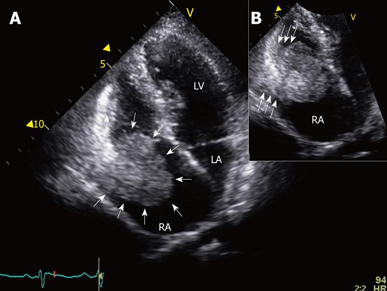
Figure 1 Transthoracic echocardiography.
A: A mobile, round, spherical, and echogenic mass (white arrows) is seen adjacent to the lateral right atrial wall on an apical four chamber view of the transthoracic echocardiography; B: A magnified image of the right atrial mass shows a pedunculated character with a broad stalk (tris-arrows). RA: Right atrium; LA: Left atrium; LV: Left ventricle.
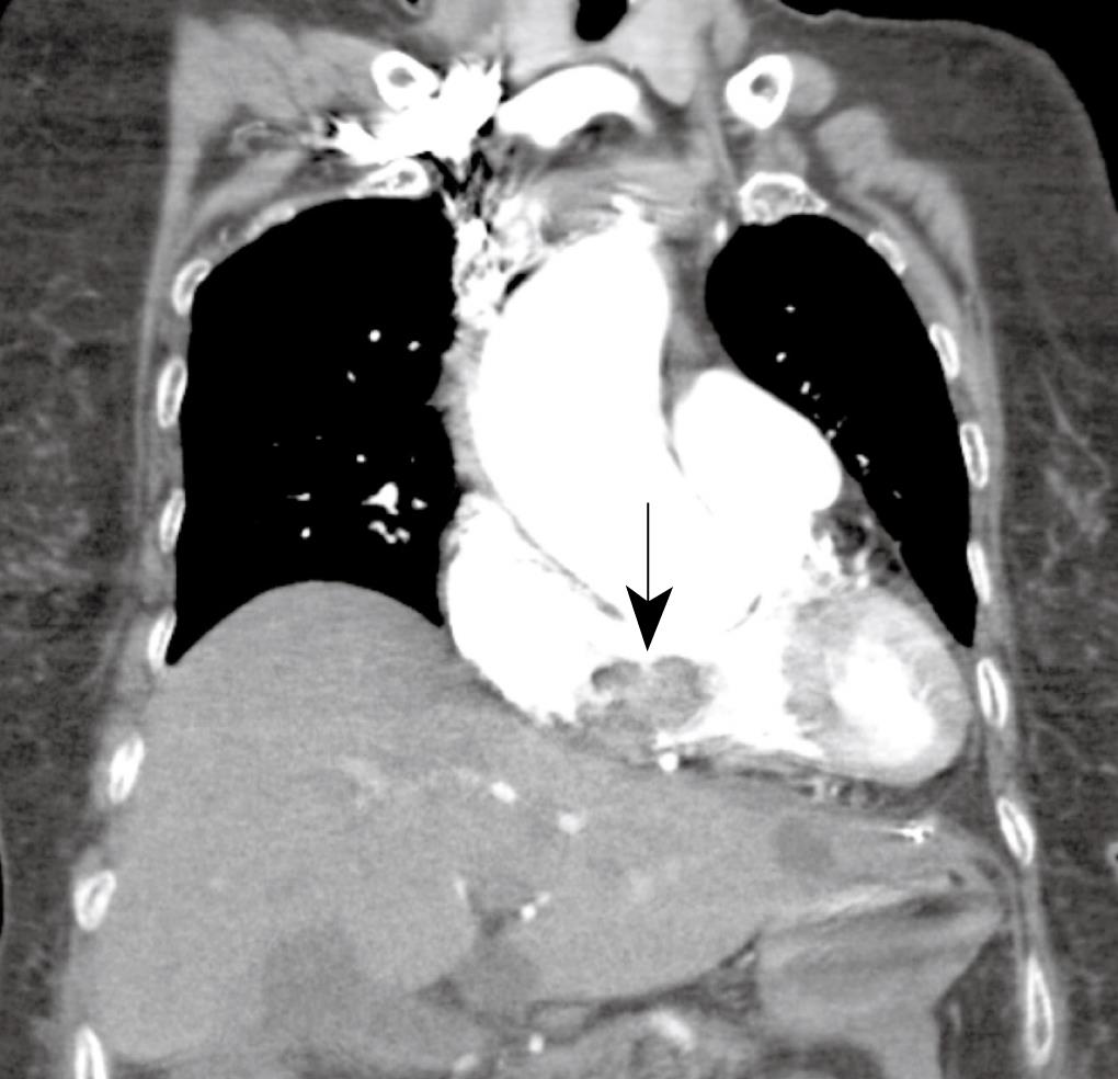
Figure 2 Computed tomography scan revealed that the mass (arrow) was located in the right atrium, obstructing the tricuspid valve opening.
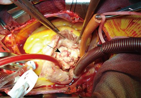
Figure 3 Operative finding.
On opening the right atrium, a large multiple lobulating mass (arrow) with a rough surface was located on the antero-inferior side of the right atrial free wall. The mass was near the atrioventricular groove, with invasion into the right atrium.
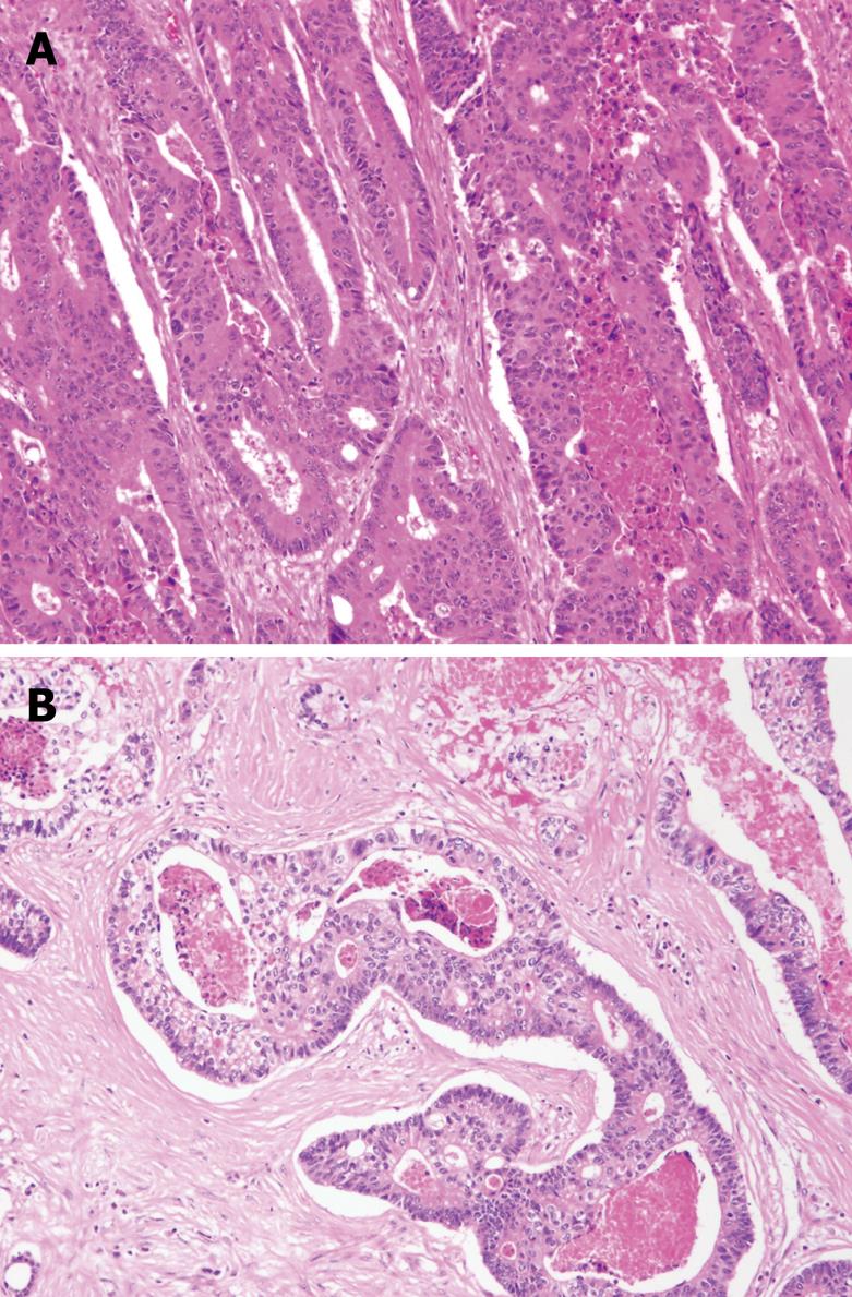
Figure 4 Microscopic findings.
A: Tall malignant columnar cells line large irregular glands; some forming a cribriform architecture containing intraluminal necrotic debris in primary colon cancer (HE, × 100); B: The cardiac mass shows similar histological findings to Figure 4A.
- Citation: Choi PW, Kim CN, Chang SH, Chang WI, Kim CY, Choi HM. Cardiac metastasis from colorectal cancer: A case report. World J Gastroenterol 2009; 15(21): 2675-2678
- URL: https://www.wjgnet.com/1007-9327/full/v15/i21/2675.htm
- DOI: https://dx.doi.org/10.3748/wjg.15.2675


