Copyright
©2008 The WJG Press and Baishideng.
World J Gastroenterol. Jan 7, 2008; 14(1): 136-139
Published online Jan 7, 2008. doi: 10.3748/wjg.14.136
Published online Jan 7, 2008. doi: 10.3748/wjg.14.136
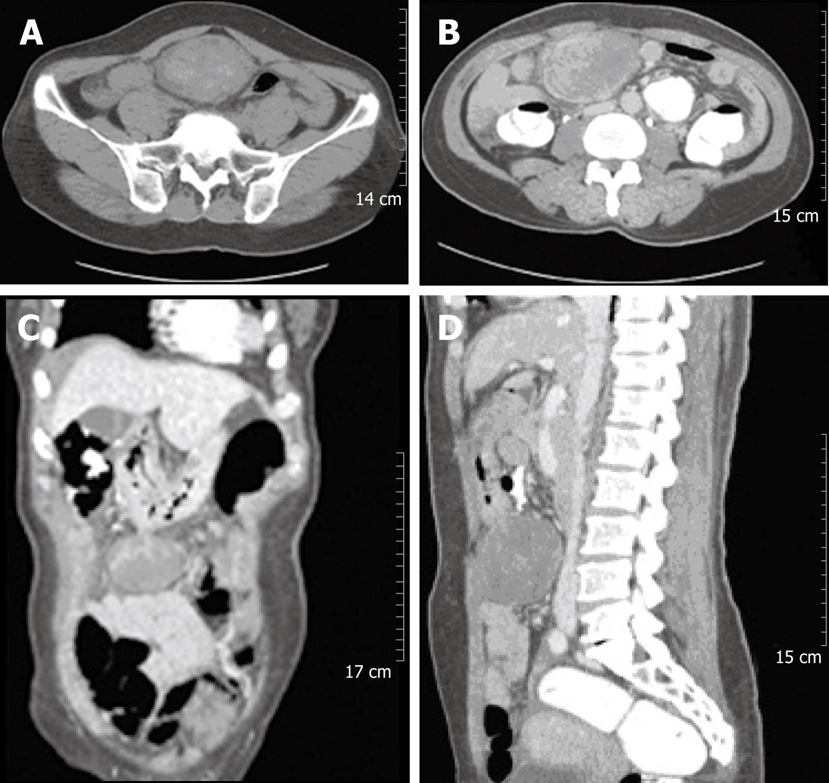
Figure 1 Abdominal CT revealed a large solid mass at the gastrocolic ligament or the gastric wall, which showed heterogeneous density on an non-enhanced image (A).
The 8 cm mass showed internally enhanced vessels on the arterial phase of CT and delayed peripheral enhancement of the mass on the venous phase (B-D).
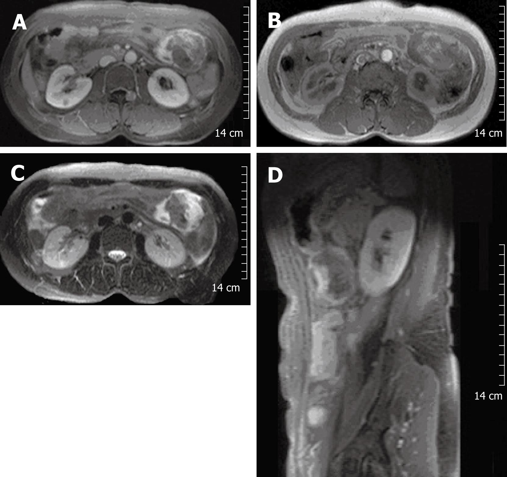
Figure 2 Contrast-enhanced MRI of the abdomen showed a mass of approximately 8 cm, seen at the left upper quadrant of the abdomen.
The margin of the mass was lobulating, and it was attached to the greater curvature of the stomach. It contained a peripheral enhanced solid portion and a central non-enhancing portion (A). Signal intensity of the central non-enhancing portion was low on T1WI (B) and T2WI (C and D), which suggested internal hemorrhage within the tumor.
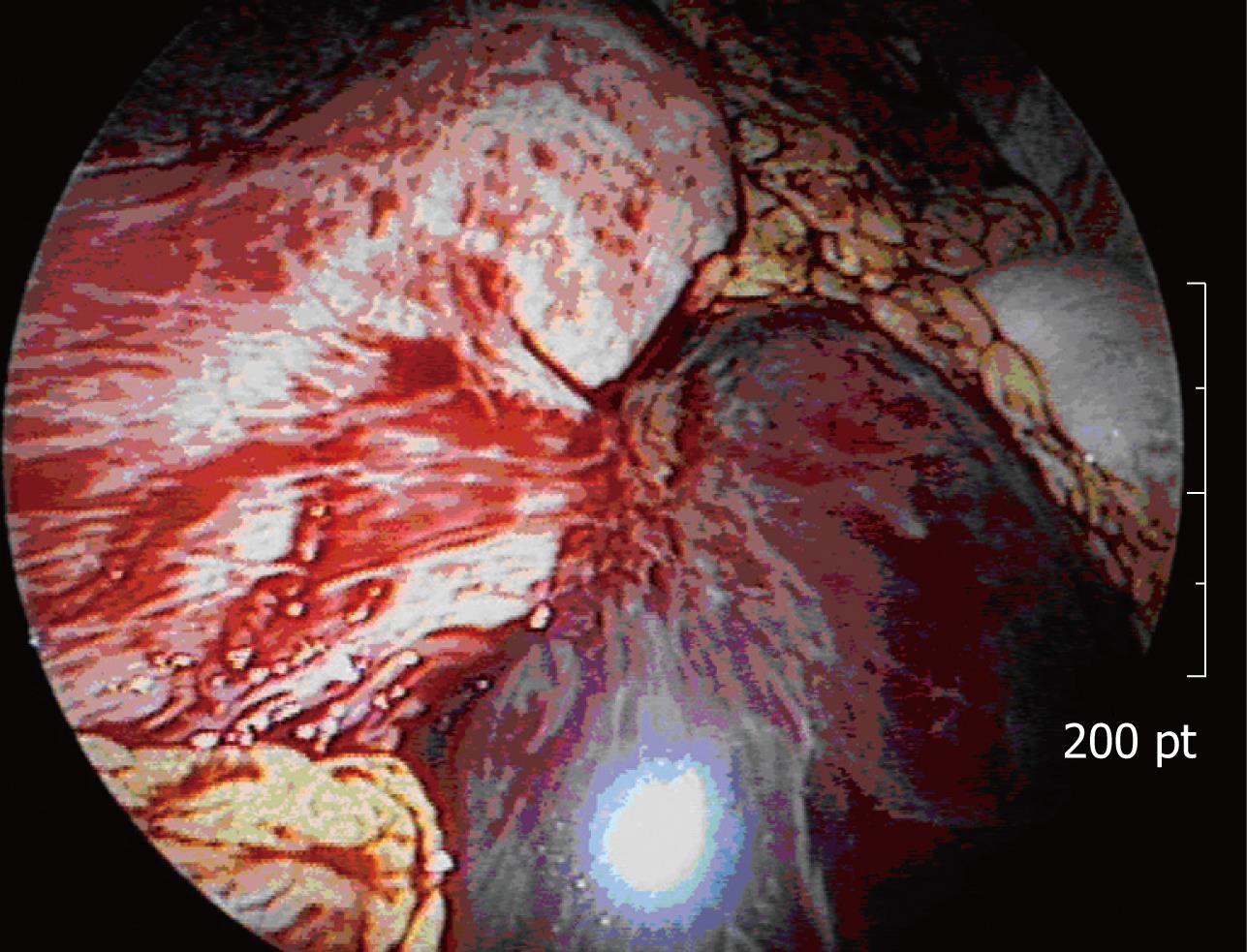
Figure 3 Laparoscopic view of the exophytic gastric mass with a large amount of intra-abdominal non-clotting hemorrhage.
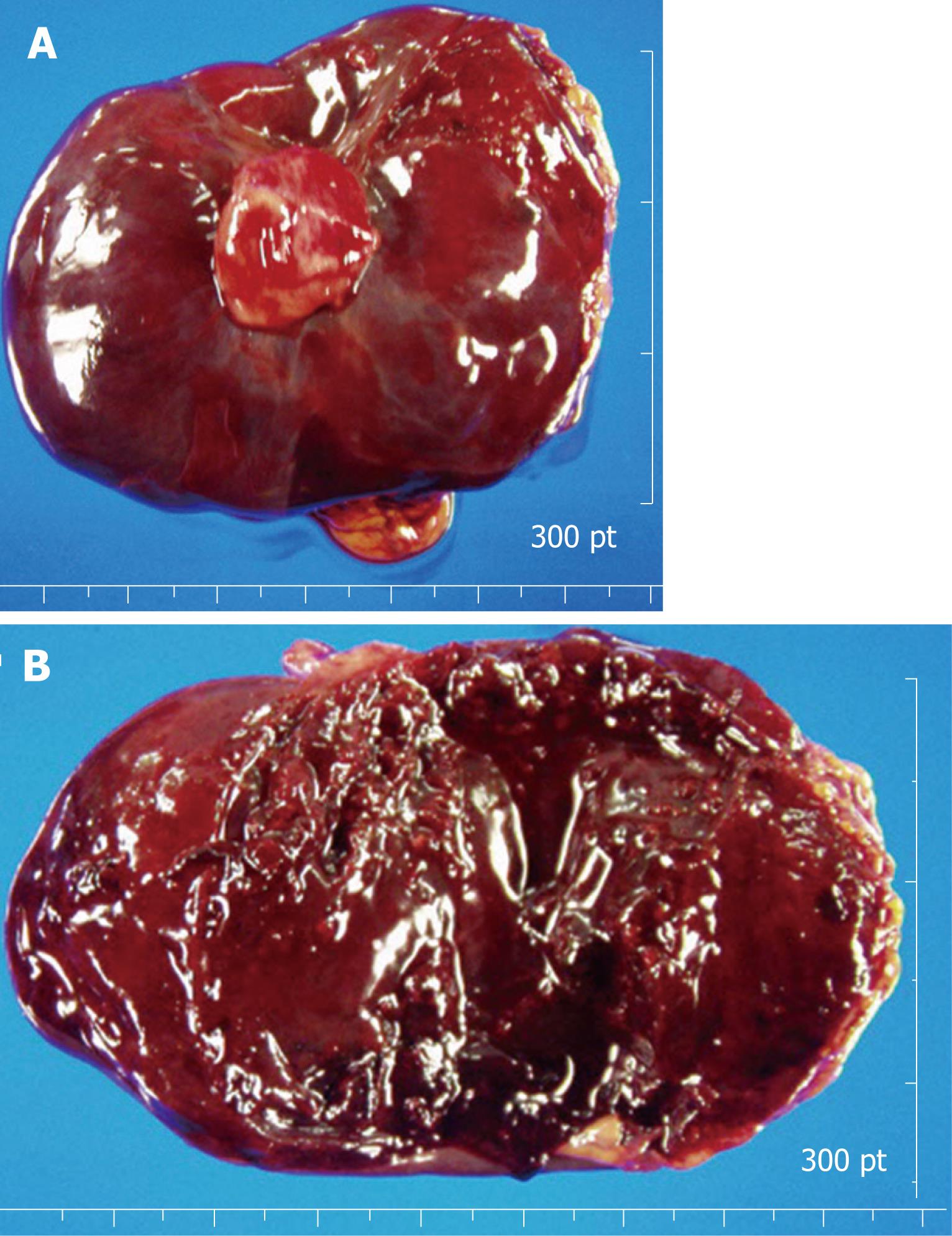
Figure 4 The external surface of a well-encapsulated lump of soft solid tumor, weighing 88.
1 g, was smooth and glistening, but showed no gastric mucosal lesion (A). Cross-sectional surface of the tumor was characterized by several amorphous fragments of parenchymal tissue, which were separated by the cystic spaces (B).
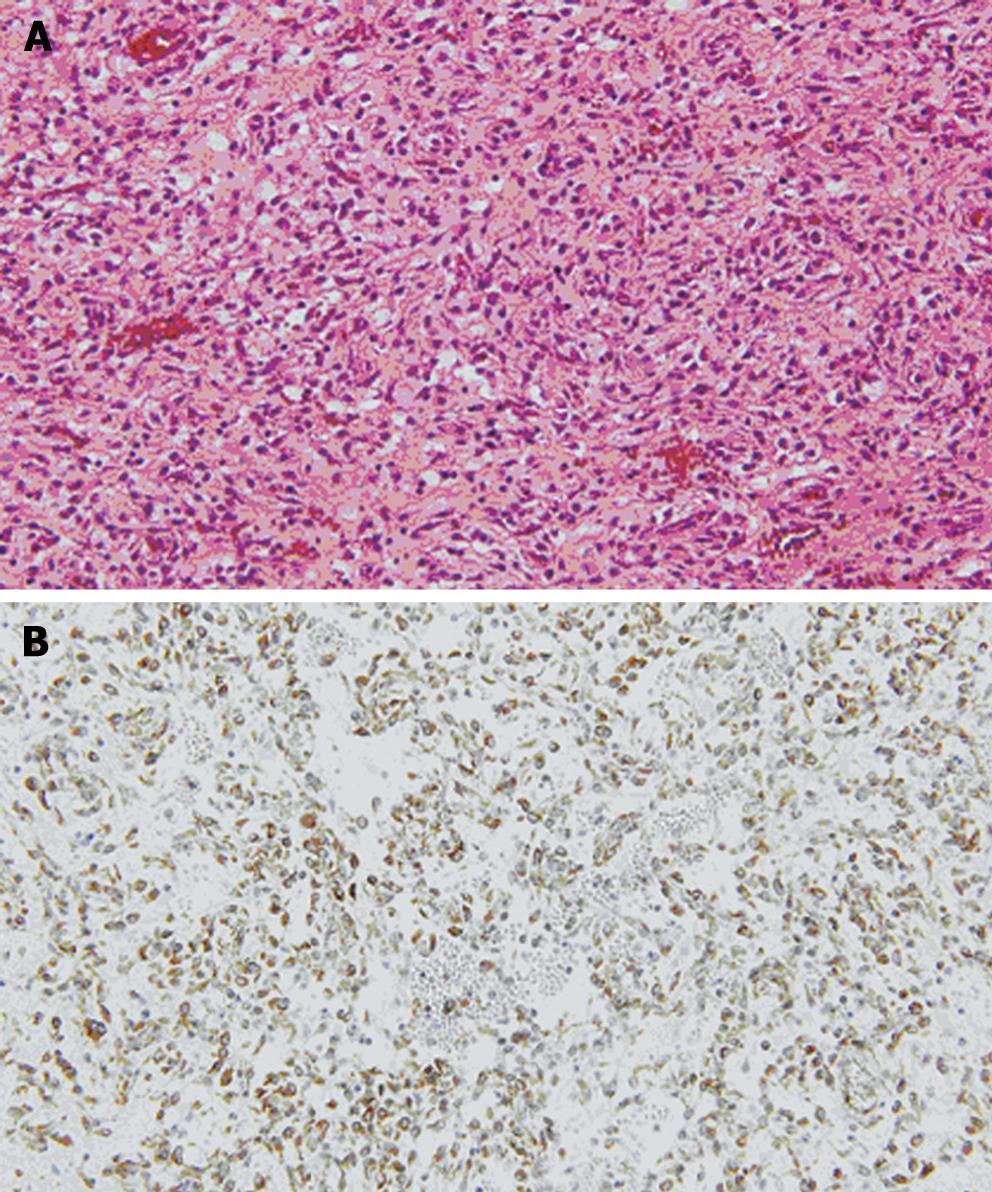
Figure 5 A: The tumor was composed of round and spindle-shaped myofibroblastic cells.
Diffusely scattered inflammatory cells and many vascular structures are seen (HE, × 400); B: The tumor cells showed positive immuno-reactivity for vimentin (× 400).
- Citation: Park SH, Kim JH, Min BW, Song TJ, Son GS, Kim SJ, Lee SW, Chung HH, Lee JH, Um JW. Exophytic inflammatory myofibroblastic tumor of the stomach in an adult woman: A rare cause of hemoperitoneum. World J Gastroenterol 2008; 14(1): 136-139
- URL: https://www.wjgnet.com/1007-9327/full/v14/i1/136.htm
- DOI: https://dx.doi.org/10.3748/wjg.14.136


