Copyright
©2008 The WJG Press and Baishideng.
World J Gastroenterol. Jan 7, 2008; 14(1): 136-139
Published online Jan 7, 2008. doi: 10.3748/wjg.14.136
Published online Jan 7, 2008. doi: 10.3748/wjg.14.136
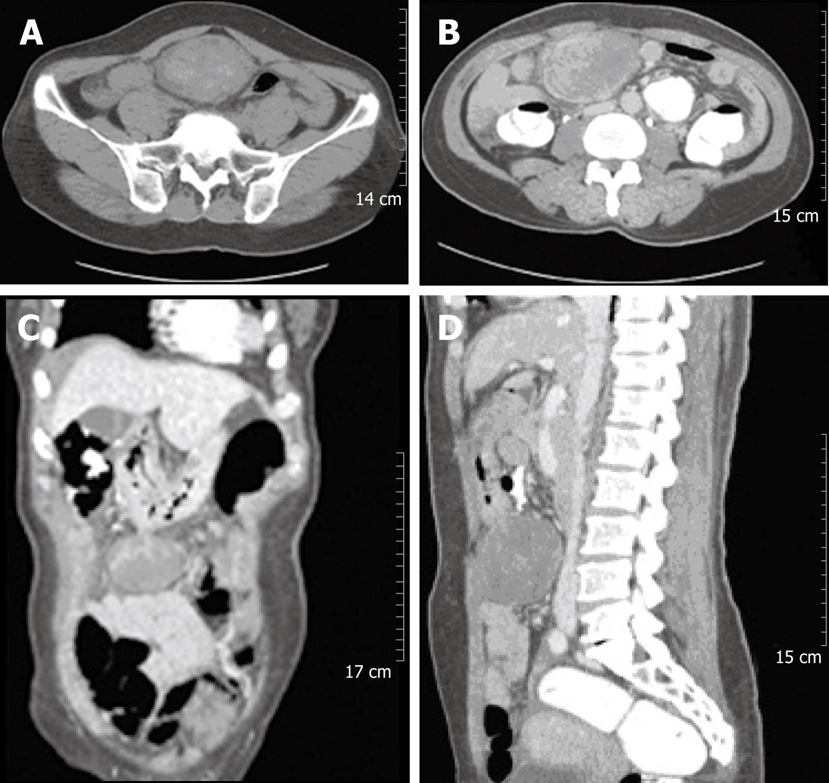
Figure 1 Abdominal CT revealed a large solid mass at the gastrocolic ligament or the gastric wall, which showed heterogeneous density on an non-enhanced image (A).
The 8 cm mass showed internally enhanced vessels on the arterial phase of CT and delayed peripheral enhancement of the mass on the venous phase (B-D).
- Citation: Park SH, Kim JH, Min BW, Song TJ, Son GS, Kim SJ, Lee SW, Chung HH, Lee JH, Um JW. Exophytic inflammatory myofibroblastic tumor of the stomach in an adult woman: A rare cause of hemoperitoneum. World J Gastroenterol 2008; 14(1): 136-139
- URL: https://www.wjgnet.com/1007-9327/full/v14/i1/136.htm
- DOI: https://dx.doi.org/10.3748/wjg.14.136


