Copyright
©2007 Baishideng Publishing Group Co.
World J Gastroenterol. Nov 28, 2007; 13(44): 5951-5953
Published online Nov 28, 2007. doi: 10.3748/wjg.v13.i44.5951
Published online Nov 28, 2007. doi: 10.3748/wjg.v13.i44.5951
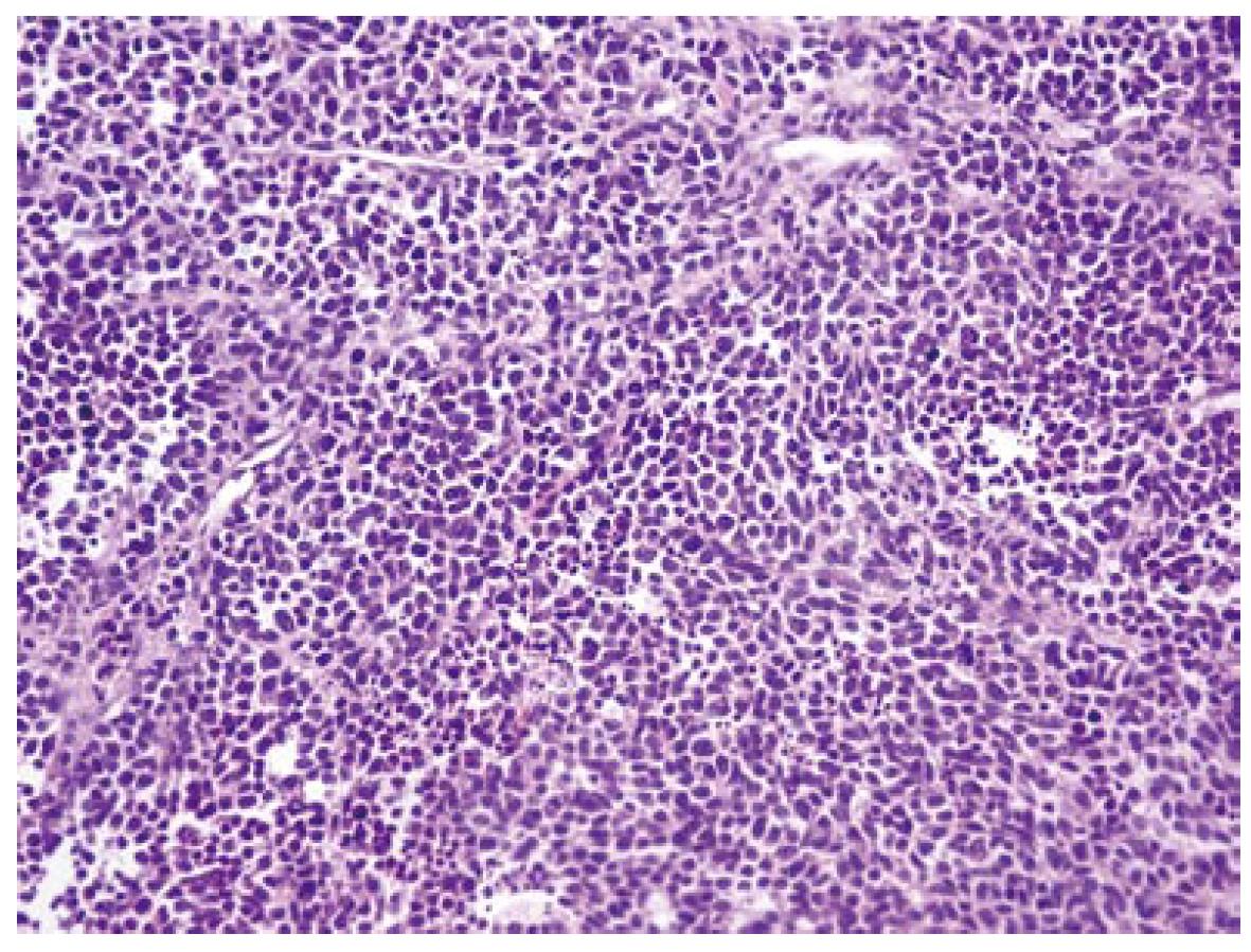
Figure 1 Histological appearance of the colorectal adenocarcinoma (HE, × 20).
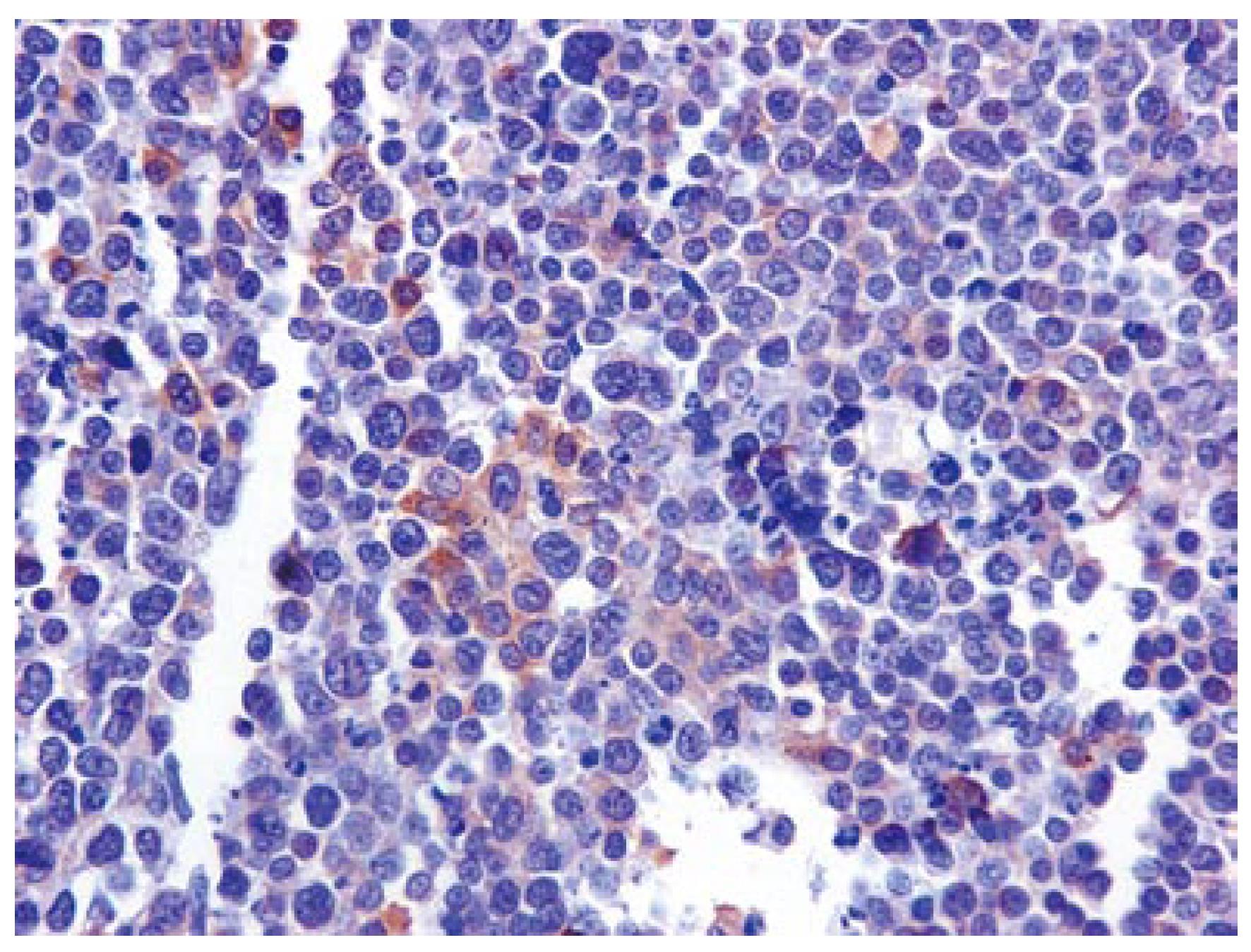
Figure 2 Scanty CD-56 immunohistochemical expression by tumor cells (× 40).
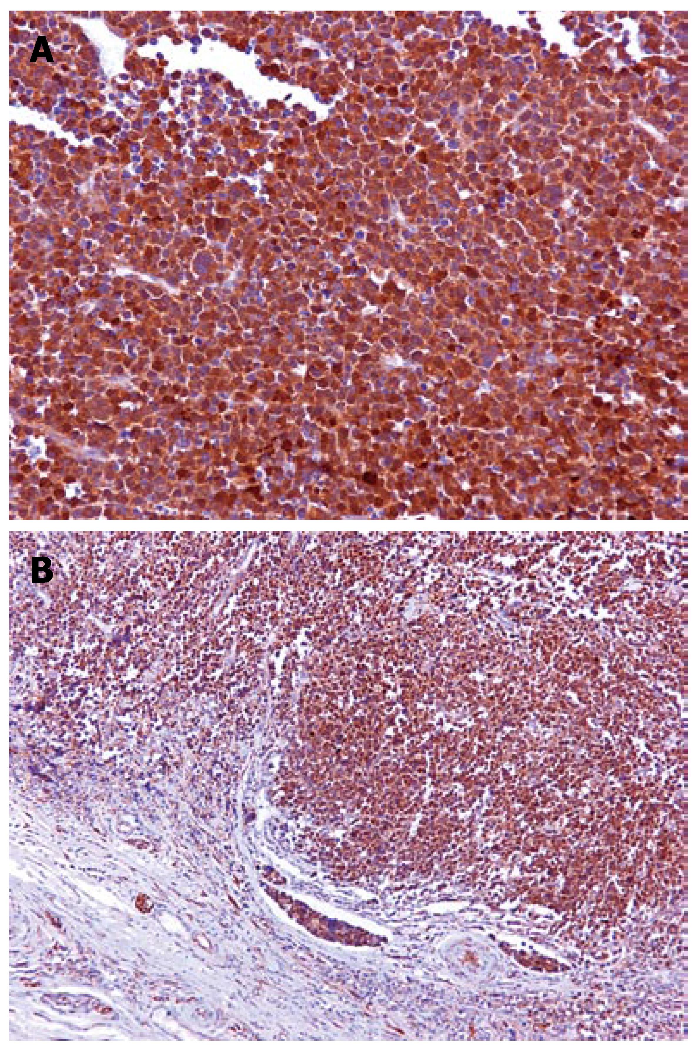
Figure 3 A: Intense c-kit immunolabeling (× 20); B: intense nuclear cytoplasmic immunolabeling for c-kit protein (× 10).
- Citation: Giannopoulos A, Papaconstantinou I, Alexandrou P, Petrou A, Papalambros A, Felekouras E, Papalambros E. Poorly differentiated carcinoma of the rectum with aberrant immunophenotype: A case report. World J Gastroenterol 2007; 13(44): 5951-5953
- URL: https://www.wjgnet.com/1007-9327/full/v13/i44/5951.htm
- DOI: https://dx.doi.org/10.3748/wjg.v13.i44.5951


