Copyright
©2006 Baishideng Publishing Group Co.
World J Gastroenterol. May 28, 2006; 12(20): 3186-3195
Published online May 28, 2006. doi: 10.3748/wjg.v12.i20.3186
Published online May 28, 2006. doi: 10.3748/wjg.v12.i20.3186
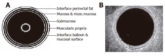
Figure 1 Rectal wall anatomy.
A: Schematic diagram of ERUS image; B: Actual image of normal ERUS[3].
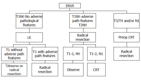
Figure 2 Treatment algorithm for patients with rectal cancer and no evidence of distant metastases.
LE: local excision; CRT: chemoradiation therapy. Observation following a LE of a T1 adenocarcinoma, even with good pathological features, may result in 20% local recurrence at 10 years.
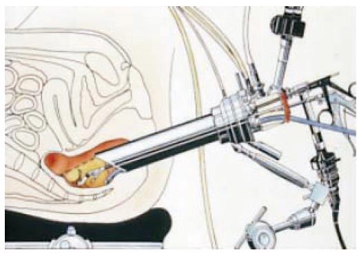
Figure 3 Specialized equipment in use for the performance of TEM[90].
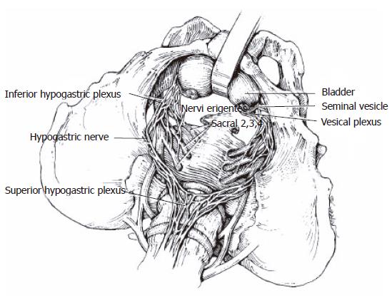
Figure 4 Diagram of pelvic autonomic nerve anatomy[73].
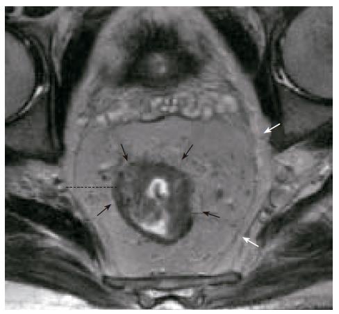
Figure 5 MRI of rectal cancer with demonstration of circumferential resection margin (CRM)[6]; Black arrows: rectal cancer; White arrows: mesorectal fascia; Dashed line: CRM, which is defined as the shortest distance from rectal cancer to the lateral resection margin of the mesorectum.
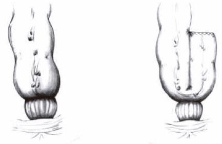
Figure 6 Illustrated comparison of a straight coloanal anastomosis and a coloanal anastomosis with a colonic J-pouch[74].
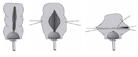
Figure 7 Technique of construction of a stapled coloanal anastomosis with a transverse coloplasty pouch.
Alternatively, the pouch may be hand-sewn to the anal canal[77].
- Citation: Balch GC, Meo AD, Guillem JG. Modern management of rectal cancer: A 2006 update. World J Gastroenterol 2006; 12(20): 3186-3195
- URL: https://www.wjgnet.com/1007-9327/full/v12/i20/3186.htm
- DOI: https://dx.doi.org/10.3748/wjg.v12.i20.3186


