Copyright
©2005 Baishideng Publishing Group Inc.
World J Gastroenterol. Feb 28, 2005; 11(8): 1248-1249
Published online Feb 28, 2005. doi: 10.3748/wjg.v11.i8.1248
Published online Feb 28, 2005. doi: 10.3748/wjg.v11.i8.1248
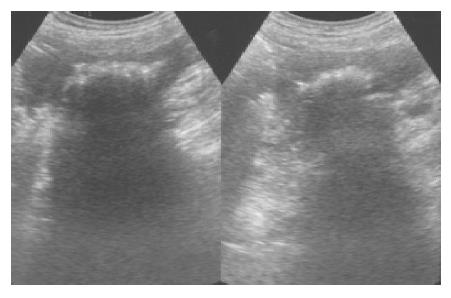
Figure 1 Right intercostal sonogram obtained 4 mo after surgery show a cystic mass with irregular internal components.
Posteriorly, the mass has strong acoustic shadowing.
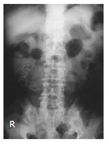
Figure 2 Plain roentgenogram of abdomen after abdominal sonogram.
Notice serpentine linear opacity over the right portion of abdomen, some mottled hyperdensities are adjacent to this linear opacity- all diagnostic features of a retained gauze.
- Citation: Shyung LR, Chang WH, Lin SC, Shih SC, Kao CR, Chou SY. Report of gossypiboma from the standpoint in medicine and law. World J Gastroenterol 2005; 11(8): 1248-1249
- URL: https://www.wjgnet.com/1007-9327/full/v11/i8/1248.htm
- DOI: https://dx.doi.org/10.3748/wjg.v11.i8.1248


