Copyright
©The Author(s) 2004.
World J Gastroenterol. Feb 1, 2004; 10(3): 348-351
Published online Feb 1, 2004. doi: 10.3748/wjg.v10.i3.348
Published online Feb 1, 2004. doi: 10.3748/wjg.v10.i3.348
Math 1 Math(A1).
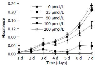
Figure 1 Proliferation kinetics of SGC-7901 cells treated with β-ionone at various concentrations.
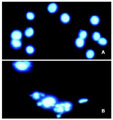
Figure 2 β-ionone-induced apoptosis in SGC-7901 cells stained with Hoechst-33258 (× 400).
A: control SGC-7901 cells, B: ex-perimental cells treated with β-ionone showing nuclear shrink-age or fragmentation.
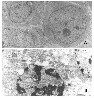
Figure 3 Ultrastructural changes of SGC-7901 cells treated with 100 μmol/L β-ionone for 24 h (× 15000).
A: SGC-7901 cells in the control group, B: experimental SGC-7901 cells treated with β-ionone showing early changes of apoptosis in which nuclear chromatin condensation and cell shrinkage were observed.
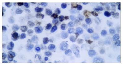
Figure 4 Apoptotic cells induced by β-ionone (200 μmol/L 48 h) in SGC-7901 cells detected TUNEL assay (× 400).
- Citation: Liu JR, Chen BQ, Yang BF, Dong HW, Sun CH, Wang Q, Song G, Song YQ. Apoptosis of human gastric adenocarcinoma cells induced by β-ionone. World J Gastroenterol 2004; 10(3): 348-351
- URL: https://www.wjgnet.com/1007-9327/full/v10/i3/348.htm
- DOI: https://dx.doi.org/10.3748/wjg.v10.i3.348


