Copyright
©The Author(s) 2004.
World J Gastroenterol. Jun 1, 2004; 10(11): 1678-1681
Published online Jun 1, 2004. doi: 10.3748/wjg.v10.i11.1678
Published online Jun 1, 2004. doi: 10.3748/wjg.v10.i11.1678
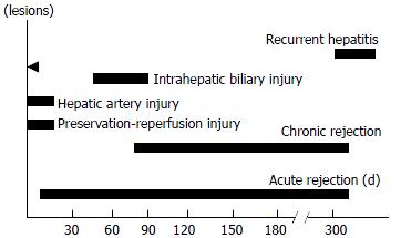
Figure 1 Time features of different complications in allograft liver biopsy.
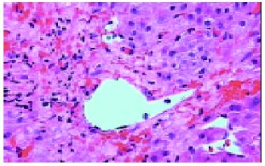
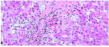
Figure 2 Hepatocytes necrosis and bleeding surrounding central vein caused by HAT 12 d after transplantation, HE × 200.
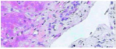
Figure 3 Bile duct damage with inflammation 80 d after transplantation.
HE × 200.
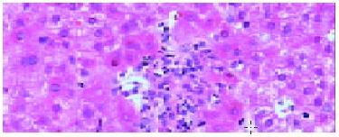
Figure 4 CMV infection in peripheral blood and micro-ab-scess without inclusion bodies in liver cells 1 mo after transplantation.
HE × 200.
Figure 5 Bile duct loss in portal tract compatible with early chronic rejection 3 mo after transplantation.
HE × 200.
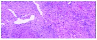
Figure 6 Fibrosis in portal area with lymphocyte infiltration and interface hepatitis in patient with recurrent hepatitis B 300 d post-transplantation, HE × 100.
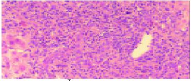
Figure 7 Acute rejection and mixed inflammatory cells in portal area with bile duct infiltration and venous endothelial inflammation 7 d post-transplantation.
HE × 100.
- Citation: Yu YY, Ji J, Zhou GW, Shen BY, Chen H, Yan JQ, Peng CH, Li HW. Liver biopsy in evaluation of complications following liver transplantation. World J Gastroenterol 2004; 10(11): 1678-1681
- URL: https://www.wjgnet.com/1007-9327/full/v10/i11/1678.htm
- DOI: https://dx.doi.org/10.3748/wjg.v10.i11.1678


