Copyright
©The Author(s) 2004.
World J Gastroenterol. Jun 1, 2004; 10(11): 1569-1573
Published online Jun 1, 2004. doi: 10.3748/wjg.v10.i11.1569
Published online Jun 1, 2004. doi: 10.3748/wjg.v10.i11.1569
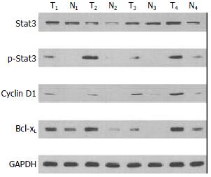
Figure 1 Expressions of Stat3, p-Stat3, cyclin D1, and Bcl-xL in colorectal carcinoma.
Lysates were made as described under Materials and Methods. GAPDH represents the internal pro-tein control. Elevated levels of Stat3, p-Stat3 (Tyr-705), cyclin D1, and Bcl-xL in tumor (T) tissues were compared to adjacent normal mucosae (N).
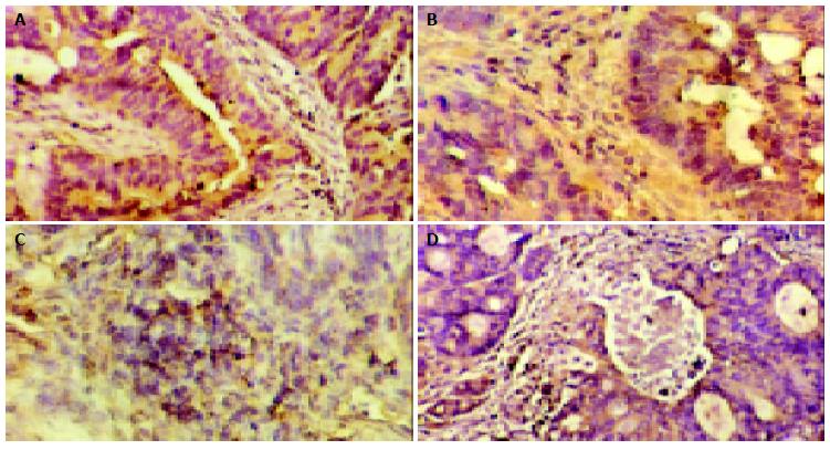
Figure 2 Expressions of Stat3, p-Stat3, cyclin D1, and Bcl-xL in colorectal carcinoma.
A: Cytoplasmic staining of Stat3 in CRC (original magnification × 200) ; B: Nuclear staining of p-Stat3 in CRC (original magnification × 200) ; C: Nuclear staining of cyclin D1 in CRC (original magnification × 200) ; D: Cytoplasmic staining of Bcl-xL (original magnification × 200).
- Citation: Ma XT, Wang S, Ye YJ, Du RY, Cui ZR, Somsouk M. Constitutive activation of Stat3 signaling pathway in human colorectal carcinoma. World J Gastroenterol 2004; 10(11): 1569-1573
- URL: https://www.wjgnet.com/1007-9327/full/v10/i11/1569.htm
- DOI: https://dx.doi.org/10.3748/wjg.v10.i11.1569


