Copyright
©The Author(s) 2021.
Artif Intell Med Imaging. Apr 28, 2021; 2(2): 32-36
Published online Apr 28, 2021. doi: 10.35711/aimi.v2.i2.32
Published online Apr 28, 2021. doi: 10.35711/aimi.v2.i2.32
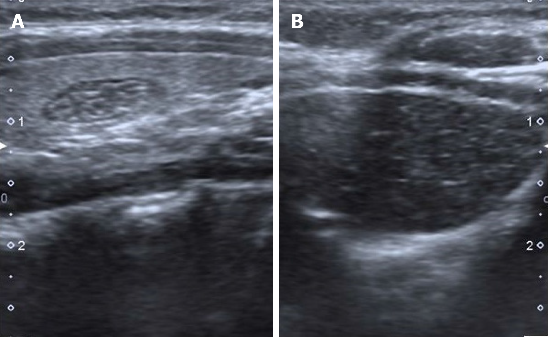
Figure 1 Eight-year-old asymptomatic girl.
Longitudinal sonographic images obtained with 7 MHz linear transducer. A: Intrathyroidal ectopic thymus with typical ultrasonographic findings; hypoechoic, fusiform appearance with linear and punctate echogenic focil; B: The resemblance with mediastinal thymic tissue can be seen easily.
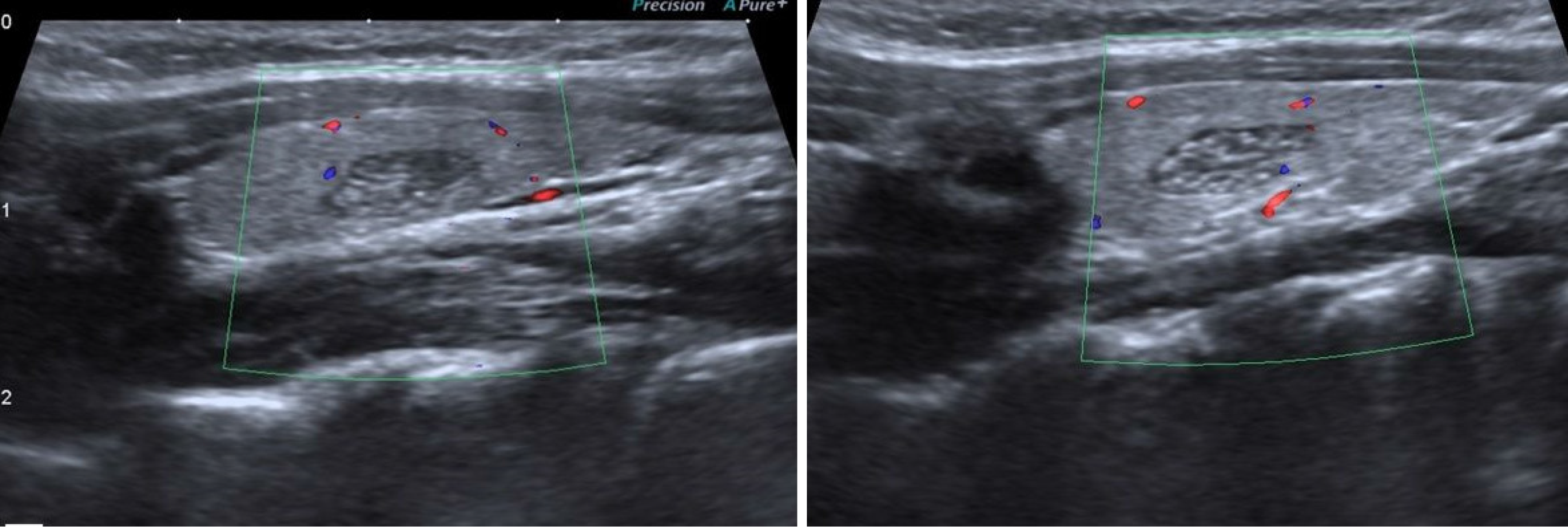
Figure 2 Eight-year-old asymptomatic girl.
Longitudinal sonographic images obtained with 7 MHz linear transducer. Both intrathyroidal ectopic thymus cases are hypo vascular in comparison within the surrounding thyroid parenchyma.
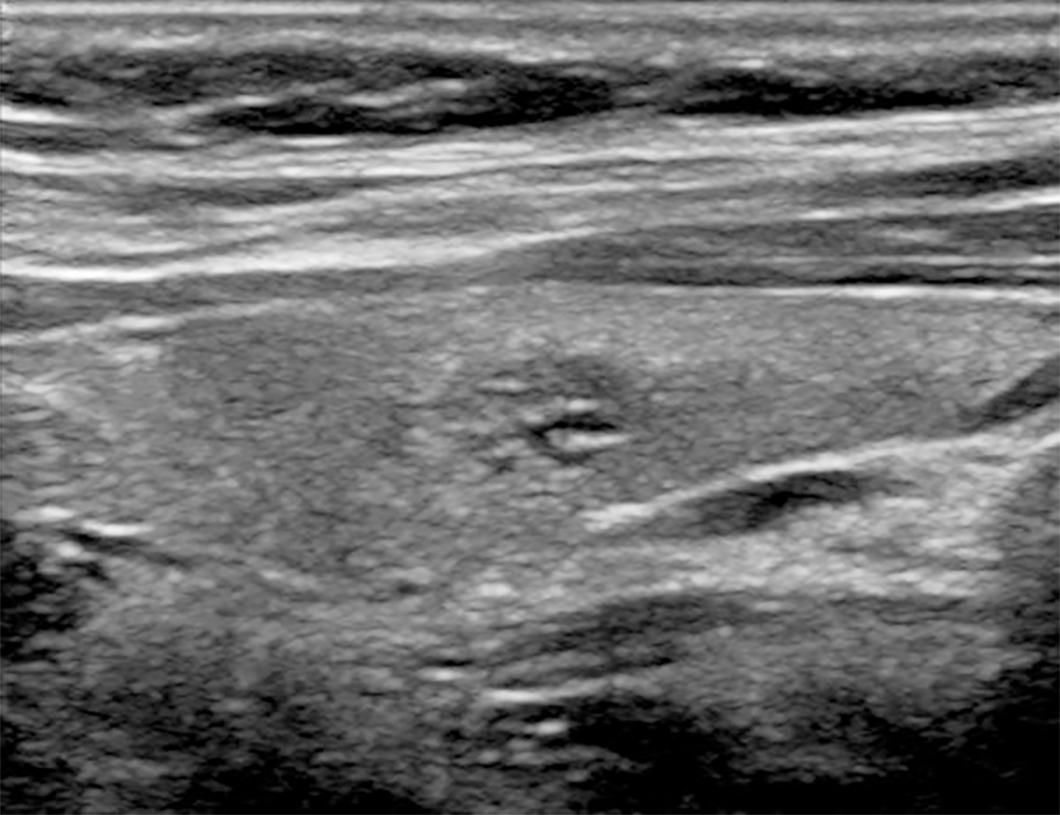
Figure 3 Nine-year-old male with hypothyroidism symptoms.
Longitudinal sonographic images obtained with 7 MHz linear transducer. A thyroid nodule presenting with a similar appearance with intrathyroidal ectopic thymus tissue.
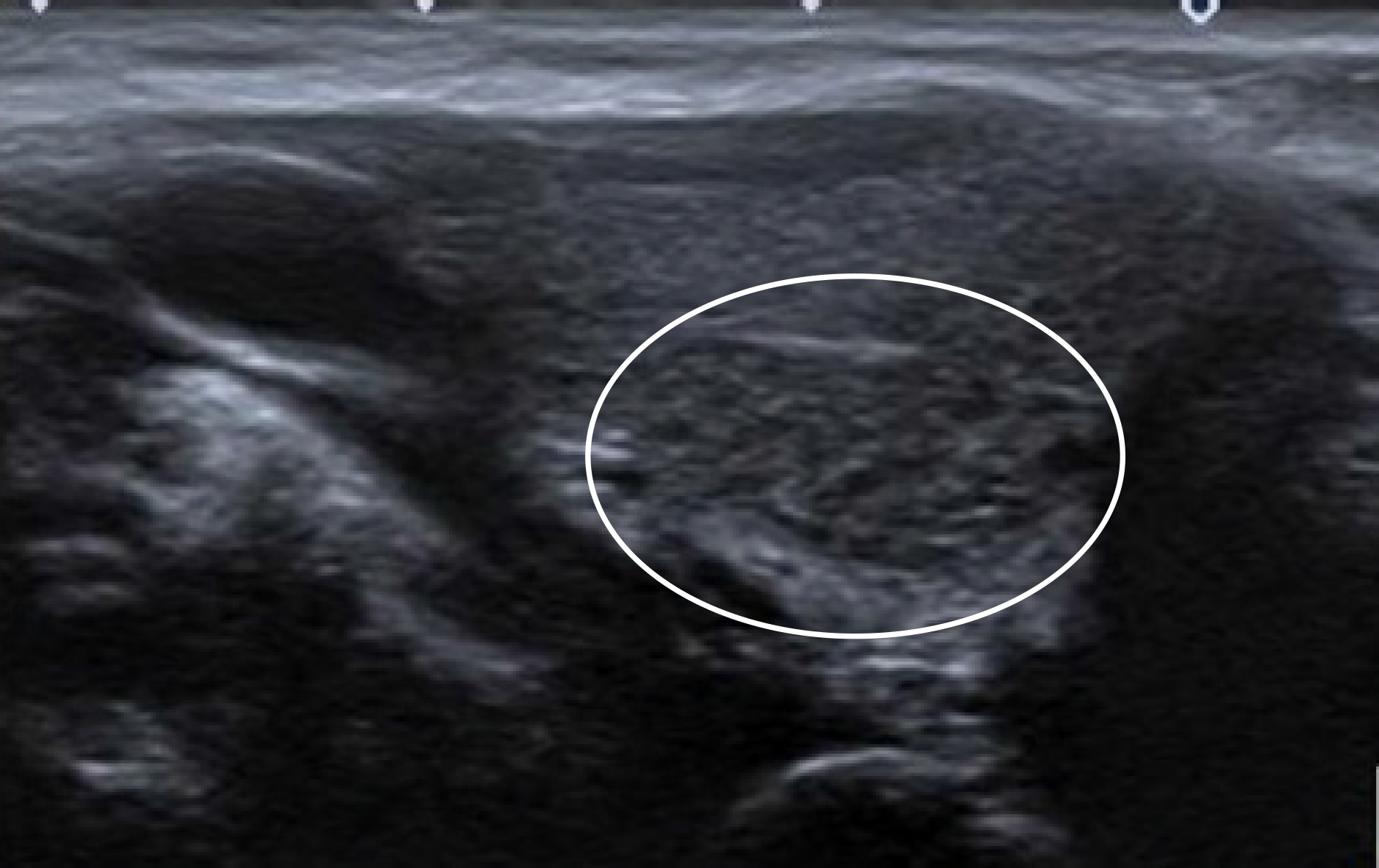
Figure 4 Ten-year-old male with recurrent cough.
Longitudinal sonographic images obtained with 7 MHz linear transducer. Abutting type intrathyroidal ectopic thymus. The lesion is located at the lower half of the gland, it has unclear margins. This appearance can be confused with focal thyroiditis.
- Citation: Karavas E, Tokur O, Aydın S, Gokharman D, Uner C. Intrathyroidal ectopic thymus: Ultrasonographic features and differential diagnosis. Artif Intell Med Imaging 2021; 2(2): 32-36
- URL: https://www.wjgnet.com/2644-3260/full/v2/i2/32.htm
- DOI: https://dx.doi.org/10.35711/aimi.v2.i2.32


