Copyright
©The Author(s) 2021.
Artif Intell Med Imaging. Apr 28, 2021; 2(2): 32-36
Published online Apr 28, 2021. doi: 10.35711/aimi.v2.i2.32
Published online Apr 28, 2021. doi: 10.35711/aimi.v2.i2.32
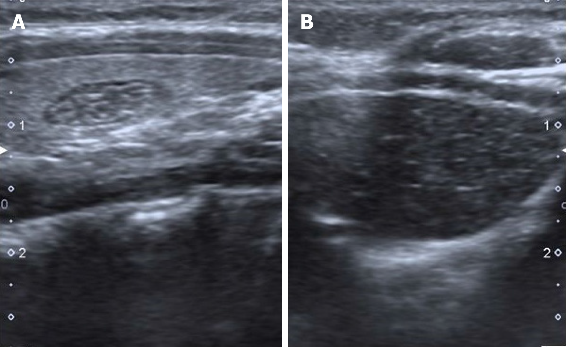
Figure 1 Eight-year-old asymptomatic girl.
Longitudinal sonographic images obtained with 7 MHz linear transducer. A: Intrathyroidal ectopic thymus with typical ultrasonographic findings; hypoechoic, fusiform appearance with linear and punctate echogenic focil; B: The resemblance with mediastinal thymic tissue can be seen easily.
- Citation: Karavas E, Tokur O, Aydın S, Gokharman D, Uner C. Intrathyroidal ectopic thymus: Ultrasonographic features and differential diagnosis. Artif Intell Med Imaging 2021; 2(2): 32-36
- URL: https://www.wjgnet.com/2644-3260/full/v2/i2/32.htm
- DOI: https://dx.doi.org/10.35711/aimi.v2.i2.32


