Copyright
©The Author(s) 2020.
World J Meta-Anal. Aug 28, 2020; 8(4): 285-291
Published online Aug 28, 2020. doi: 10.13105/wjma.v8.i4.285
Published online Aug 28, 2020. doi: 10.13105/wjma.v8.i4.285
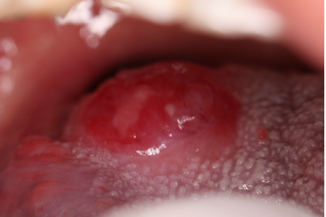
Figure 1 Clinical photograph showing the gross appearance of tumor in the right root of tongue.
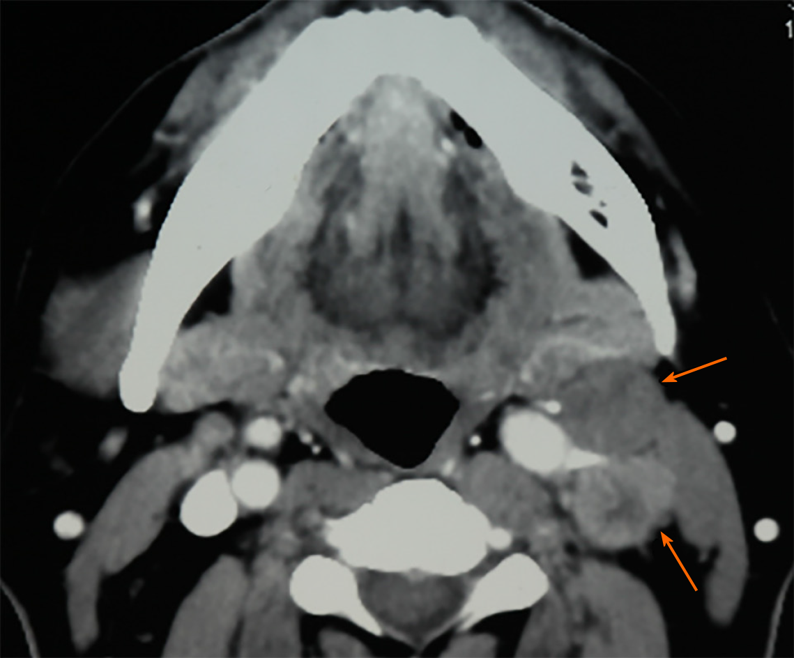
Figure 2 Preoperative computerized tomography scan showing contralateral cervical lymph nodes enlargement (orange arrows).
Figure 3 Microscopically, round and spindly small cells with ovoid- or spindle-shaped nuclei, fine granular chromatin, inconspicuous nucleoli and scant cytoplasm formed broad nests, sheets or cord shapes.
A: The tumor cells were located in the fiber and striated muscle tissues below the mucous membrane, arranging in nest, sheet and cord shapes (× 100); B: The tumor cells were found to infiltrate the fiber and striated muscle tissues, secondary inflammatory reactions were also found in the interstitial fiber, vascular tissue and nerve tissue, and infiltrating lymphocytes and neutrophils were present (× 200); C: The nucleolus of the tumor cells were unsuspicious but heteromorphosis and nuclear mitosis were obvious (× 200).
Figure 4 Immunohistochemical analysis showed positivity for (A) synaptophysin, (B) chromogranin A, and (C) cytokeratin AE1/AE3.
Magnification: × 200.
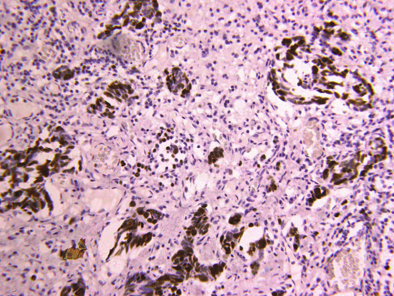
Figure 5 About 90% of the tumor cells showed Ki67+ nuclear staining, suggesting a high proliferation activity.
- Citation: Zhou Y, Zhou HC, Peng H, Zhang ZH. Primary small cell neuroendocrine carcinoma of the right posterior tongue. World J Meta-Anal 2020; 8(4): 285-291
- URL: https://www.wjgnet.com/2308-3840/full/v8/i4/285.htm
- DOI: https://dx.doi.org/10.13105/wjma.v8.i4.285


