
















Copyright: ©Author(s) 2026.
World J Clin Cases. Sep 26, 2026; 14(27): 126027
Published online Sep 26, 2026. doi: 10.12998/wjcc.126027
Published online Sep 26, 2026. doi: 10.12998/wjcc.126027
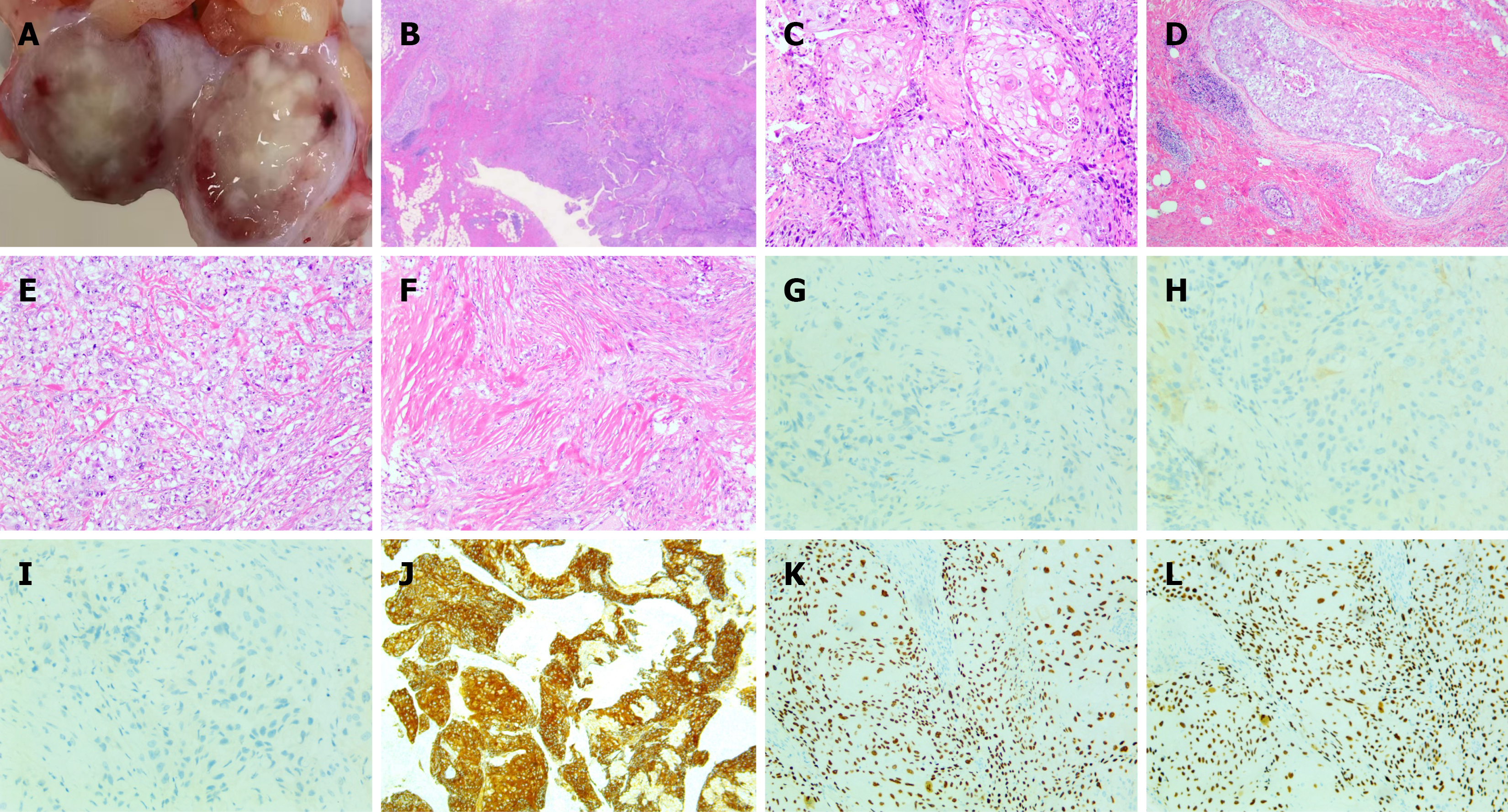
Figure 1 Tumor and histopathological features.
A: Gross specimen obtained after segmental resection of the breast mass, showing the cut surface of the lesion after central incision; B: Histopathological features of mixed metaplastic breast cancer; C: Histopathological features of squamous cell carcinoma (× 100); D: Histopathological features of high-grade ductal carcinoma in situ (× 100); E: Histopathological features of invasive ductal carcinoma (× 100); F: Histopathological features of spindle cell carcinoma (× 100); G: Immunohistochemical staining showing negative expression of estrogen receptor in breast cancer tissue (× 200); H: Immunohistochemical staining showing negative HER2 expression in breast cancer tissue (× 200); I: Immunohistochemical staining showing negative PR expression in breast cancer tissue (× 200); J: Immunohistochemical staining showing positive CK5/6 expression in breast cancer tissue (× 20); K: Immunohistochemical staining revealed positive expression of P40 in breast cancer tissues (× 20); L: Immunohistochemical staining revealed positive expression of P63 in breast cancer tissues (× 20).
- Citation: Zhao TJ, Chen SL, Zhou YL, Jin ZJ, Huang GS. Multicomponent metaplastic breast cancer with four histological components: A case report. World J Clin Cases 2026; 14(27): 126027
- URL: https://www.wjgnet.com/2307-8960/full/v14/i27/126027.htm
- DOI: https://dx.doi.org/10.12998/wjcc.126027