
















Copyright: ©Author(s) 2026.
World J Clin Cases. Sep 6, 2026; 14(25): 125804
Published online Sep 6, 2026. doi: 10.12998/wjcc.125804
Published online Sep 6, 2026. doi: 10.12998/wjcc.125804
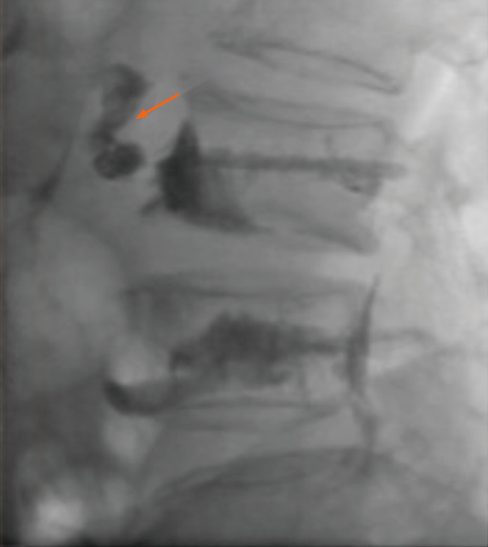
Figure 1 Lateral fluoroscopic view of the lumbar spine obtained during vertebroplasty.
The orange arrow indicates anterior extravasation of polymethylmethacrylate beyond the anterior cortex of the vertebral body, suggestive of cortical breach with leakage into the prevertebral region.
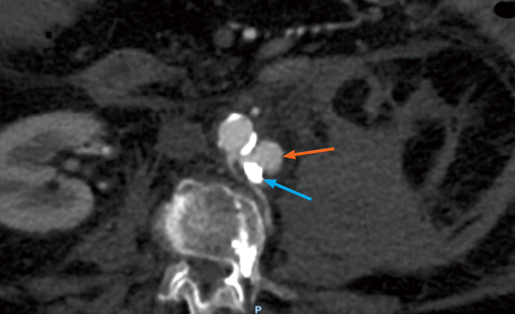
Figure 2 Axial contrast-enhanced computed tomography angiography demonstrating abdominal aortic pseudoaneurysm after vertebroplasty.
The blue arrow indicates migration of polymethylmethacrylate anterior to the vertebral body, and the orange arrow indicates the abdominal aortic pseudoaneurysm.
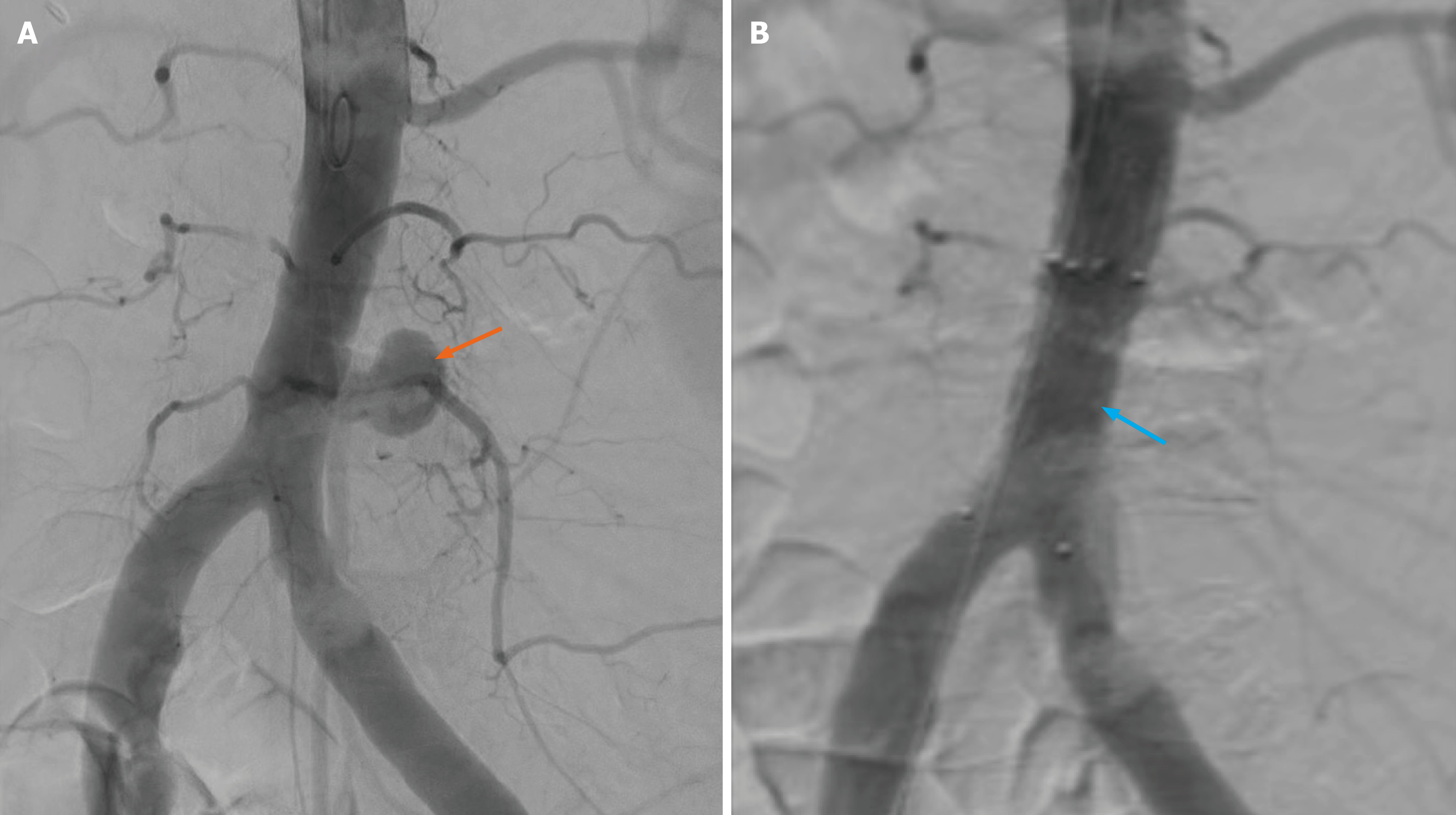
Figure 3 Aortic angiography before and after endovascular stent-graft placement.
A: Abdominal aortic pseudoaneurysm (orange arrow); B: Complete exclusion of the pseudoaneurysm, with no evidence of contrast extravasation after stent-graft deployment (blue arrow).
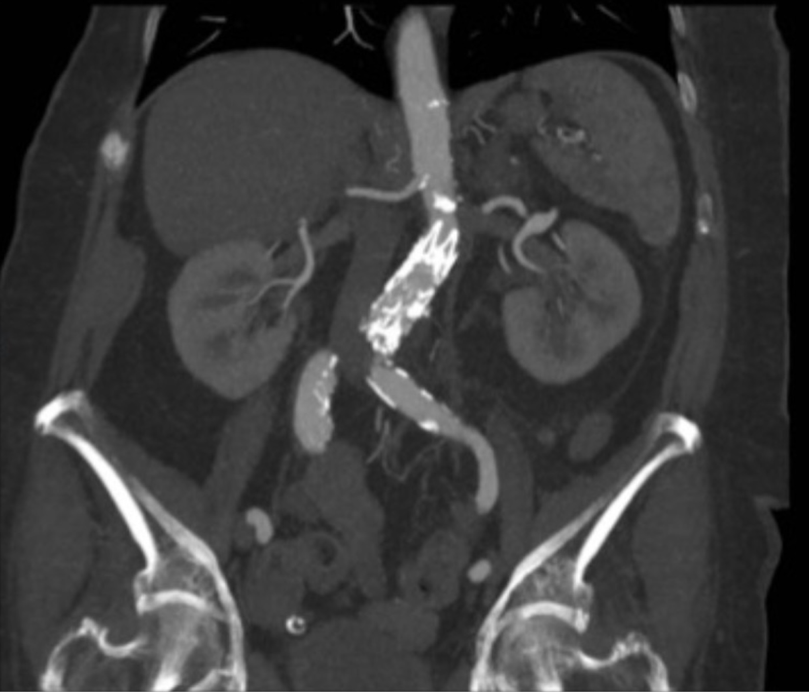
Figure 4 Six-month follow-up computed tomography angiography after endovascular stent-graft placement.
Contrast-enhanced computed tomography angiography demonstrates stable stent positioning, without endoleak, thrombosis, or migration.
- Citation: Dorobat B, Musa A, Radu RA. Abdominal aortic pseudoaneurysm as a complication of percutaneous vertebroplasty: A case report. World J Clin Cases 2026; 14(25): 125804
- URL: https://www.wjgnet.com/2307-8960/full/v14/i25/125804.htm
- DOI: https://dx.doi.org/10.12998/wjcc.125804