
















Copyright: ©Author(s) 2026.
World J Transl Med. Nov 28, 2026; 12(4): 124522
Published online Nov 28, 2026. doi: 10.5528/wjtm.124522
Published online Nov 28, 2026. doi: 10.5528/wjtm.124522
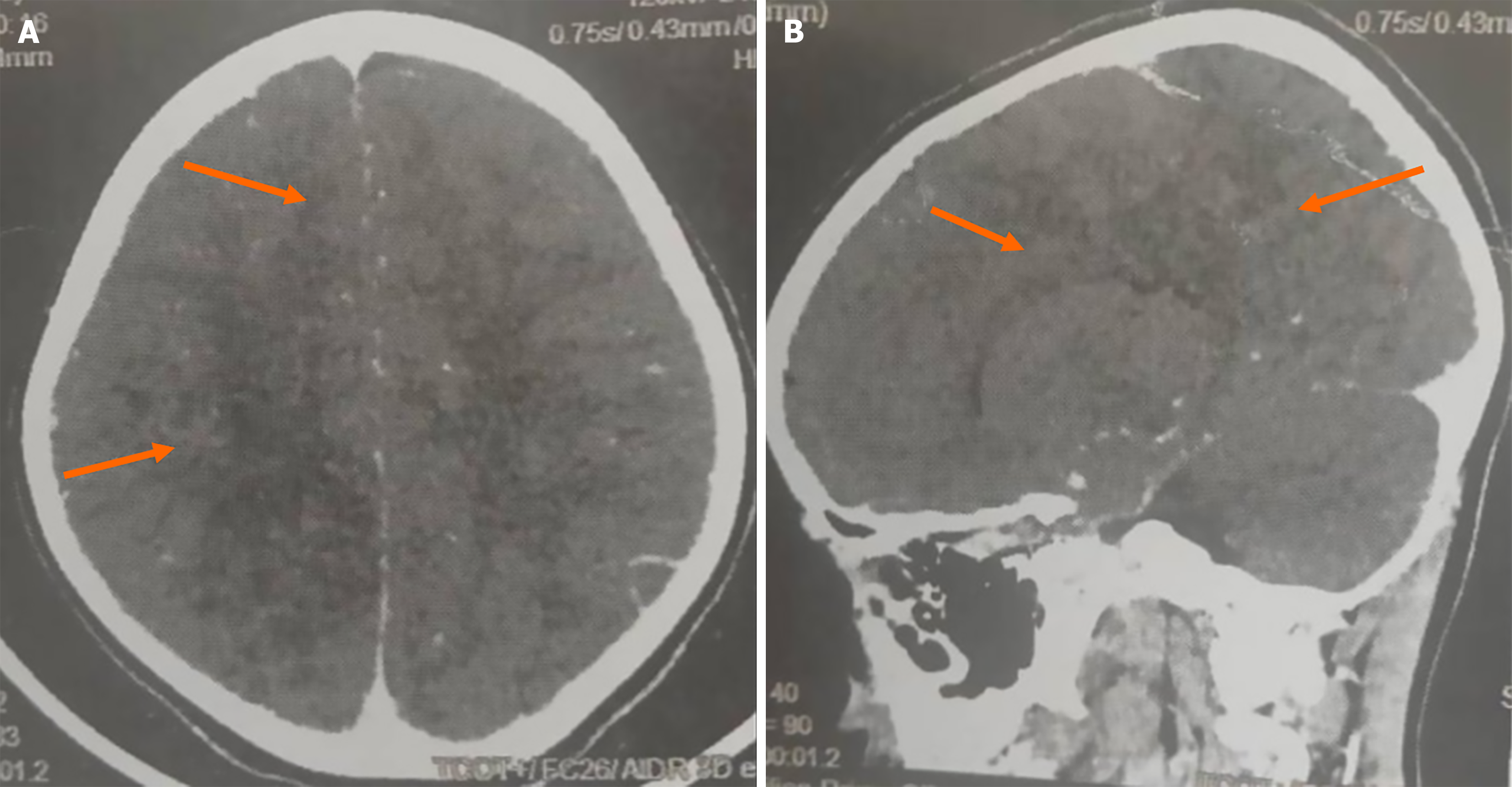
Figure 1 Brain computed tomography scans of patient, an 8-year-old girl with sickle cell disease homozygous SS.
A and B: Axial (A) and sagittal (B) views demonstrated a subacute ischemic infarction involving the territories of the right anterior cerebral artery and right middle cerebral artery. Orange arrows indicate the hypodense area corresponding to the subacute infarction.
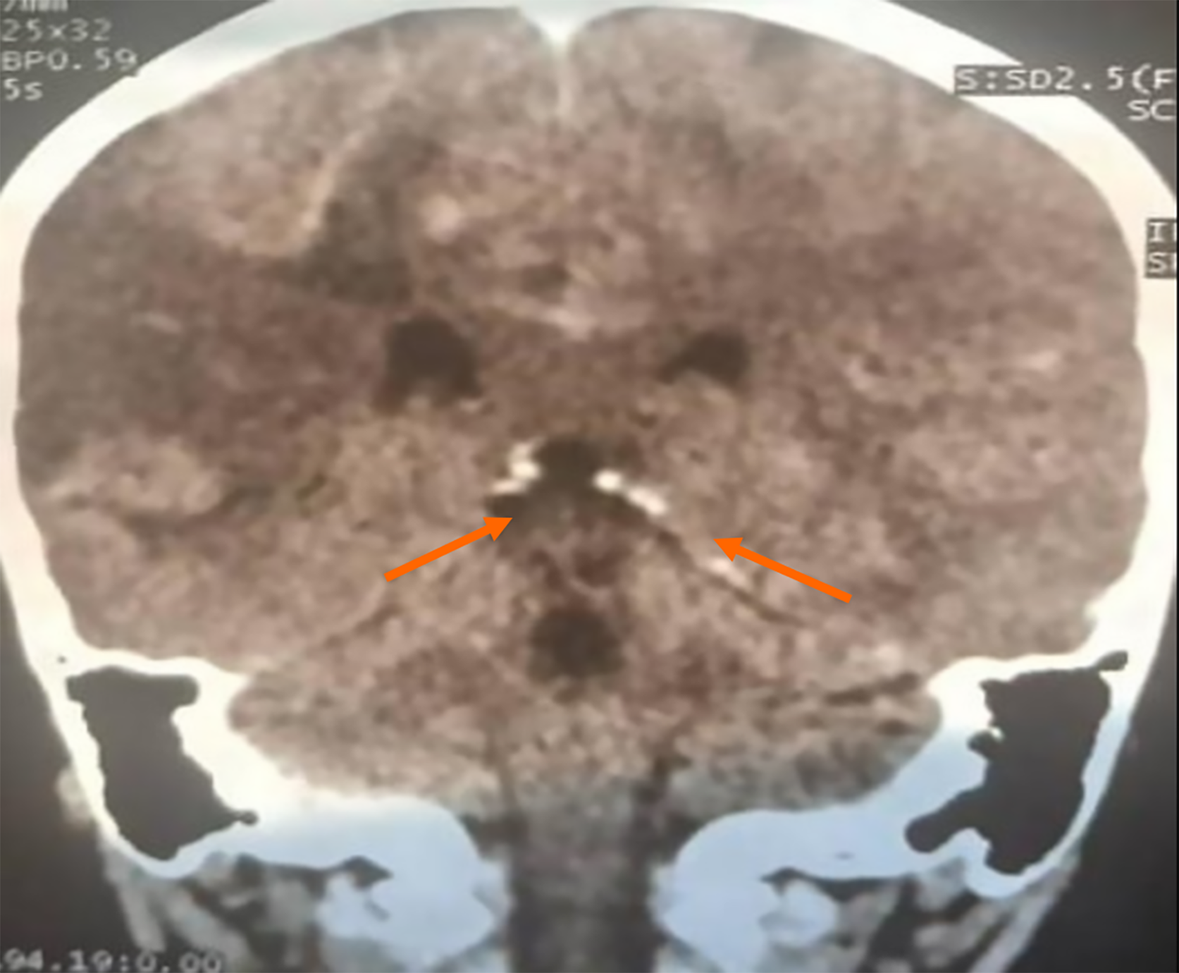
Figure 2 Brain computed tomography scan (coronal view).
Computed tomography performed on day 4 of intensive care unit hospitalization demonstrated diffuse subarachnoid hemorrhage, diffuse cerebral edema, and an approximately 8-mm right subfalcine herniation. Orange arrows indicate the hyperdense subarachnoid blood within the basal cisterns.
- Citation: Ahouangansi SER, Ouohi A, Koffi L, Kossonou AM, Kouamé KI, Toure WC, Bédié YV, Kakou KM, N’dah ES, Koné K, Adingra SCE, Aboudramane K, Ayé YD. Fatal hemorrhagic transformation after ischemic stroke in a child with sickle cell disease SS: A case report. World J Transl Med 2026; 12(4): 124522
- URL: https://www.wjgnet.com/2220-6132/full/v12/i4/124522.htm
- DOI: https://dx.doi.org/10.5528/wjtm.124522