Copyright
©The Author(s) 2016.
World J Nephrol. Nov 6, 2016; 5(6): 551-555
Published online Nov 6, 2016. doi: 10.5527/wjn.v5.i6.551
Published online Nov 6, 2016. doi: 10.5527/wjn.v5.i6.551
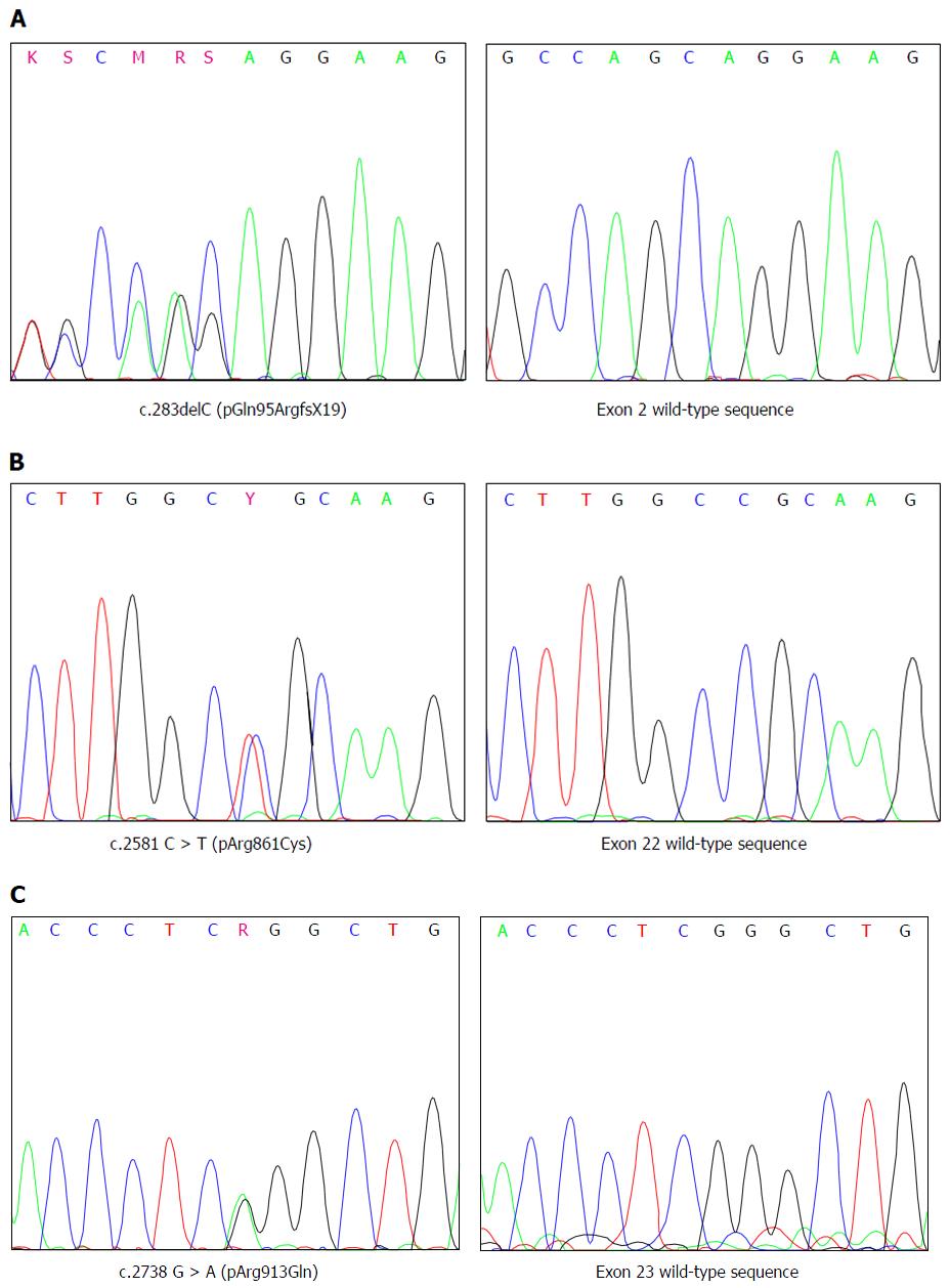
Figure 1 Electropherograms showing DNA sequences of exon 2 (A), exon 22 (B) and exon 23 (C), in the regions containing the variations detected.
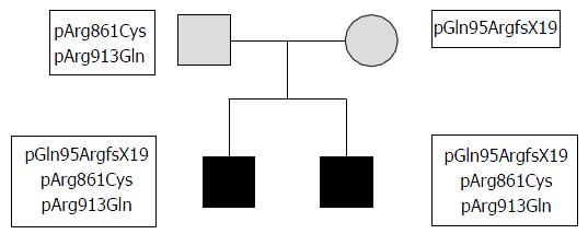
Figure 2 Pedigree of family.
Gitelman’s syndrome affected patients are colored in black, heterozygous carriers are filled in gray. Mutations and polymorphisms of subjects are reported close to their respective symbols.
- Citation: Grillone T, Menniti M, Bombardiere F, Vismara MFM, Belviso S, Fabiani F, Perrotti N, Iuliano R, Colao E. New SLC12A3 disease causative mutation of Gitelman’s syndrome. World J Nephrol 2016; 5(6): 551-555
- URL: https://www.wjgnet.com/2220-6124/full/v5/i6/551.htm
- DOI: https://dx.doi.org/10.5527/wjn.v5.i6.551


