Copyright
©2014 Baishideng Publishing Group Inc.
World J Nephrol. Nov 6, 2014; 3(4): 122-142
Published online Nov 6, 2014. doi: 10.5527/wjn.v3.i4.122
Published online Nov 6, 2014. doi: 10.5527/wjn.v3.i4.122
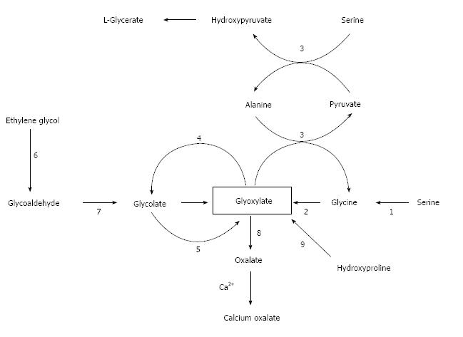
Figure 1 Biosynthesis of calcium oxalate.
Glyoxylate is the main precursor of oxalate which combines spontaneously with calcium ions to form calcium oxalate. Names of enzymes: 1, serine hydroxymethyltransferase; 2, D-amino acid oxidase; 3, alanine:glyoxylate aminotransferase (AGT); 4, glyoxylate reductase-hydroxypyruvate reductase (GRHPR); 5, glycolate oxidase; 6, alcohol dehydrogenase; 7, aldehyde dehydrogenase; 8, lactate dehydrogenase; and 9, five enzyme-catalyzed reactions. PH1 results from mutations in AGT which is a hepatic peroxisomal enzyme. PH2 results from mutations in GRHPR which is a cytosolic enzyme found in several tissues, but primarily the liver. PH3 results from defects in the hepatic mitochondrial enzyme 4-hydroxy-2-oxoglutarate (HOG) aldolase which converts HOG and glyoxylate to pyruvate (reaction not shown), the last step in hydroxyproline catabolism. The reason why a deficiency of HOG aldolase activity increases oxalate production is obscure.
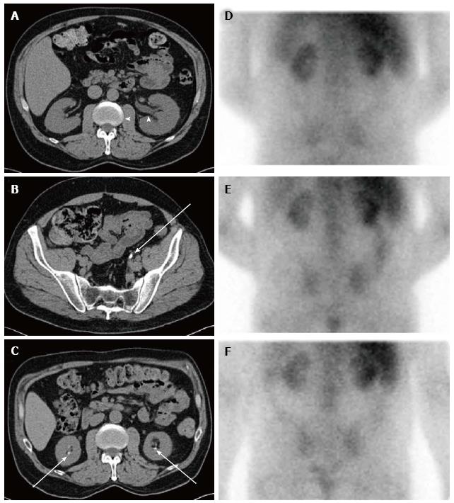
Figure 2 Sequential imaging studies of a not yet reported patient with chronic kidney disease from dietary hyperoxaluria.
Axial computed tomography (CT) images obtained two years before the hyperoxaluria diagnosis show (A) mild left hydronephrosis (arrowheads) caused by (B) a left distal ureteral calculus (arrow). Axial CT image obtained around the time of the hyperoxaluria diagnosis shows (C) bilateral nephrolithiasis (arrows). Nuclear medicine gallium-67 citrate scan images were also obtained around the time of diagnosis, including (D) 4-, (E) 24-, and (F) 48 h after administration. These show abnormal, persistent bilateral renal activity at all time points, indicative of interstitial nephritis. Gallium scanning has classically been used to distinguish acute interstitial nephritis from acute tubular necrosis and other causes of acute renal failure[216-218]. In this patient chronic interstitial nephritis associated with hyperoxaluria led to this positive scan. The patient’s diet for several years was based on nuts with estimated oxalate consumption ≥ 800 mg daily. During high oxalate intake, urine oxalate excretion was > 200 mg/24-h in several measurements obtained at serum creatinine levels > 3.5 mg/dL. After resumption of a diet low in oxalate and improvement of renal function to serum creatinine levels < 3.0 mg/dL, urine oxalate excretion decreased to normal levels.
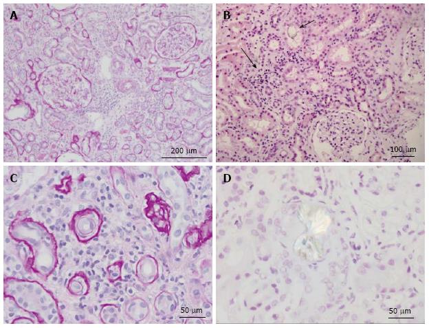
Figure 3 Renal histology in the patient depicted in Figure 2.
A: Low power view of kidney showing two complete glomeruli and expansion of the interstitium by lymphocytes and edema. Periodic acid-Schiff (PAS) stain highlights the basement membranes of the tubules and Bowman’s capsule. PAS stain; B: Low power view of renal parenchyma showing tubulointerstitial nephritis (solid arrow) and oxalate crystal within tubule (open arrow). H and E stain; C: High power view showing interstitium expanded by lymphocytic infiltrates and tubular atrophy. PAS stain; D: High power view of calcium oxalate crystal under polarized light. H and E stain.
- Citation: Glew RH, Sun Y, Horowitz BL, Konstantinov KN, Barry M, Fair JR, Massie L, Tzamaloukas AH. Nephropathy in dietary hyperoxaluria: A potentially preventable acute or chronic kidney disease. World J Nephrol 2014; 3(4): 122-142
- URL: https://www.wjgnet.com/2220-6124/full/v3/i4/122.htm
- DOI: https://dx.doi.org/10.5527/wjn.v3.i4.122


