Copyright
©2013 Baishideng Publishing Group Co.
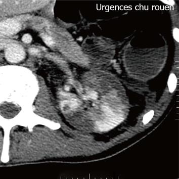
Figure 1 Contrast-enhanced helical computer tomography shows multiple demarcated areas of decreased enhancement representing focal renal infarctions.
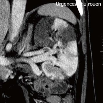
Figure 2 Axial section at the level of renal hila demonstrates a filling defect within the left renal artery due to a thrombus.
- Citation: Lemaitre C, Iwanicki-Caron I, Vecchi CD, Bertiaux-Vandaële N, Savoye G. Acute renal artery occlusion following infliximab infusion. World J Nephrol 2013; 2(3): 90-93
- URL: https://www.wjgnet.com/2220-6124/full/v2/i3/90.htm
- DOI: https://dx.doi.org/10.5527/wjn.v2.i3.90


