Copyright
©The Author(s) 2017.
World J Transplant. Feb 24, 2017; 7(1): 94-97
Published online Feb 24, 2017. doi: 10.5500/wjt.v7.i1.94
Published online Feb 24, 2017. doi: 10.5500/wjt.v7.i1.94
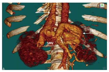
Figure 1 Active splenorenal shunt draining from the splenic vein into the left renal vein.
VCI: Vena cava inferior; SRS: Splenorenal shunt; SMV: Superior mesenteric vein.
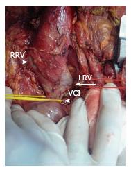
Figure 2 Anterior part of the infrahepatic vena cava was explored and dissected down to expose the bifurcation of the left renal vein.
LRV: Left renal vein; VCI: Vena cava inferior; RRV: Right renal vein.
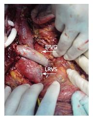
Figure 3 Right renal vein between left renal vein and graft portal vein with interposition vein graft.
IPVG: Interposition vein graft; LRVS: Left renal vein stump.
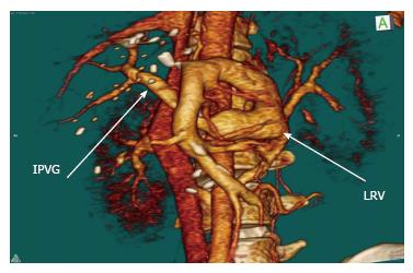
Figure 4 Computerized tomography scans visualize the patency of the right renal vein.
IPVG: Interposition vein graft; LRV: Left renal vein.
- Citation: Ozdemir F, Kutluturk K, Barut B, Abbasov P, Kutlu R, Kayaalp C, Yılmaz S. Renoportal anastomosis in living donor liver transplantation with prior proximal splenorenal shunt. World J Transplant 2017; 7(1): 94-97
- URL: https://www.wjgnet.com/2220-3230/full/v7/i1/94.htm
- DOI: https://dx.doi.org/10.5500/wjt.v7.i1.94


