Copyright
©The Author(s) 2022.
World J Transplant. Jul 18, 2022; 12(7): 204-210
Published online Jul 18, 2022. doi: 10.5500/wjt.v12.i7.204
Published online Jul 18, 2022. doi: 10.5500/wjt.v12.i7.204
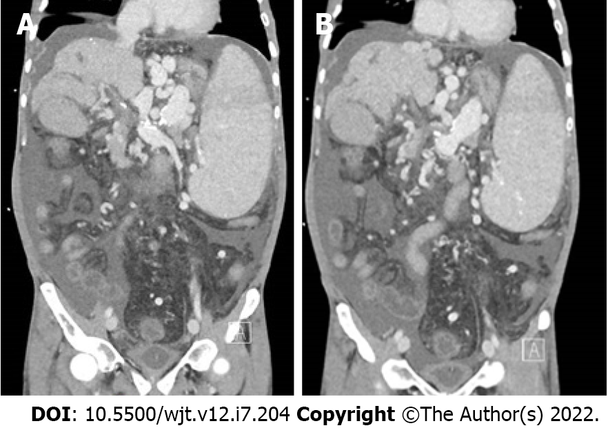
Figure 1 Preoperative abdominal computed tomography.
A: Extensive portal vein thrombosis; B: Superior mesenteric vein thrombosis.
Figure 2 Treatment imaging.
A: End-to-side portal vein-left gastric vein anastomosis upon completion; B: Postoperative Doppler sonography documenting patent anastomosis with adequate flow; C: Abdominal computed tomography showing patent portal vein-left gastric vein anastomosis.
- Citation: Gravetz A. Portal vein-variceal anastomosis for portal vein inflow reconstruction in orthotopic liver transplantation: A case report and review of literature. World J Transplant 2022; 12(7): 204-210
- URL: https://www.wjgnet.com/2220-3230/full/v12/i7/204.htm
- DOI: https://dx.doi.org/10.5500/wjt.v12.i7.204


