Copyright
©The Author(s) 2021.
World J Psychiatr. Sep 19, 2021; 11(9): 605-618
Published online Sep 19, 2021. doi: 10.5498/wjp.v11.i9.605
Published online Sep 19, 2021. doi: 10.5498/wjp.v11.i9.605
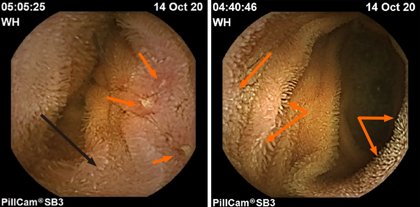
Figure 1 Pillcam images from the small bowel of a gastrointestinal-symptomatic patient with autism spectrum disorder.
Lymphangiectasia in patient with autism spectrum disorder-associated enteritis (left panel, black arrow); the patient also exhibited aphthous ulcerations (orange arrows); Jejunal lymphangiectasia (orange arrows) in adjacent mucosa of same patient (right panel).
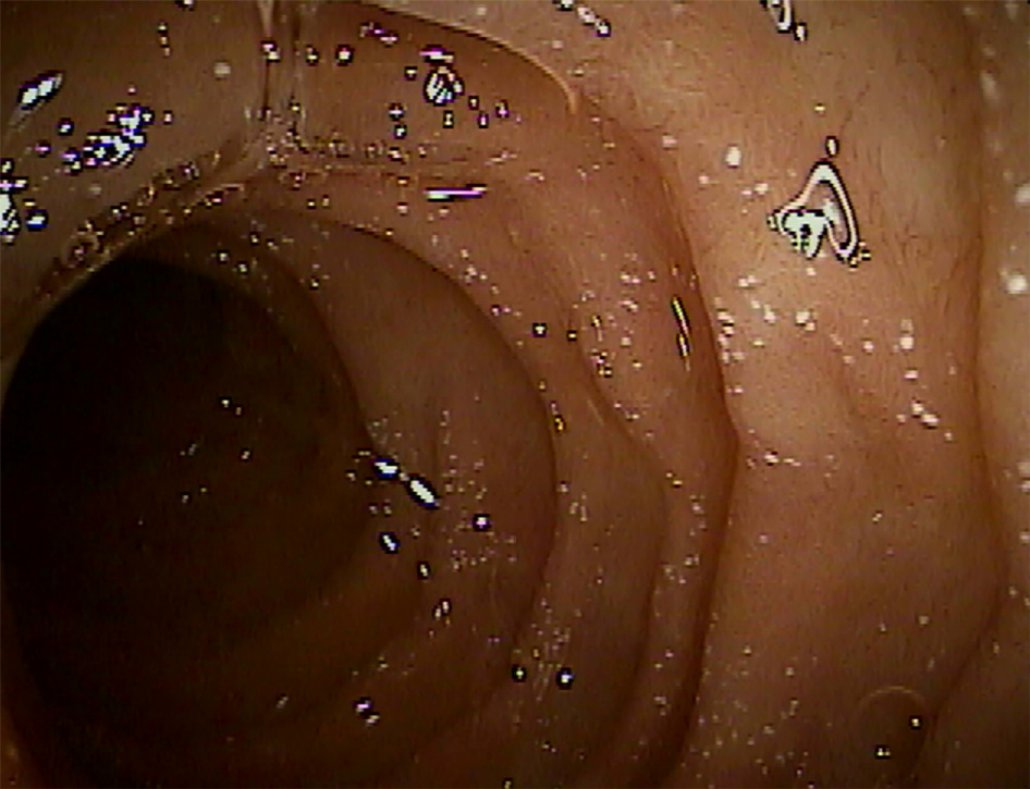
Figure 2 Endoscopic image of white spot lesions of the proximal small bowel in a gastrointestinal-symptomatic child with autism spectrum disorder.
Figure 3 Histologic staining of a mucosal biopsy specimen from the proximal small bowel of a patient with autism spectrum disorder-associated enteritis.
White spot lesions (truncated villi) can be seen in low (left); medium (middle); and high magnification (right) of the H&E-stained images.
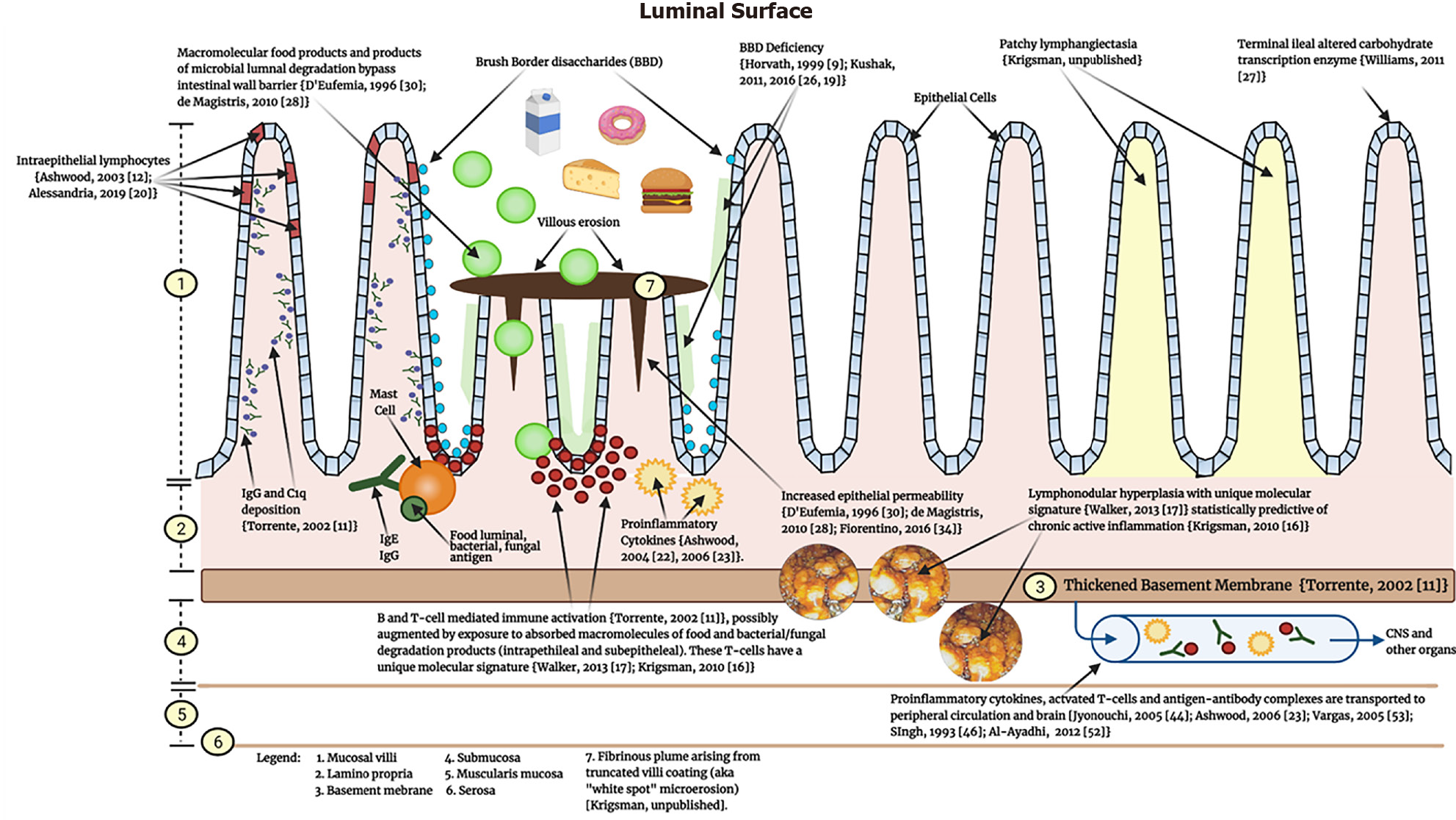
Figure 4 Schematic diagram depicting pathological findings in a portion of the gastrointestinal tract of a representative gastrointestinal-symptomatic child with autism spectrum disorder[9,11,12,16,17,19,20,22,23,26,27,28,30,34,46,52,53].
Key landmarks are identified in the legend (#1-7) and key findings are described throughout the diagram.
- Citation: Krigsman A, Walker SJ. Gastrointestinal disease in children with autism spectrum disorders: Etiology or consequence? World J Psychiatr 2021; 11(9): 605-618
- URL: https://www.wjgnet.com/2220-3206/full/v11/i9/605.htm
- DOI: https://dx.doi.org/10.5498/wjp.v11.i9.605


