Copyright
©2014 Baishideng Publishing Group Inc.
World J Clin Infect Dis. Feb 25, 2014; 4(1): 1-4
Published online Feb 25, 2014. doi: 10.5495/wjcid.v4.i1.1
Published online Feb 25, 2014. doi: 10.5495/wjcid.v4.i1.1
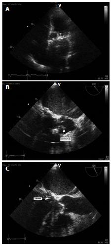
Figure 1 Transesophageal echocardiogram.
A: Transthoracic ehocardiographic image showing apical 4-chambered view showing possible vegetation on prosthetic mitral valve; B: Transesophageal echocardiogram showing sub-centimeter vegetation on the aortic valve; C: Transesophageal echocardiogram showing mobile vegetation at the prosthetic mitral valve annulus.
- Citation: Agarwal V, Parikh V, Lakhani M, De C, Motivala A, Mobarakai N. Sub-acute endocarditis by Corynebacterium straitum: An often ignored pathogen. World J Clin Infect Dis 2014; 4(1): 1-4
- URL: https://www.wjgnet.com/2220-3176/full/v4/i1/1.htm
- DOI: https://dx.doi.org/10.5495/wjcid.v4.i1.1


