Copyright
©The Author(s) 2017.
World J Exp Med. Aug 20, 2017; 7(3): 58-77
Published online Aug 20, 2017. doi: 10.5493/wjem.v7.i3.58
Published online Aug 20, 2017. doi: 10.5493/wjem.v7.i3.58
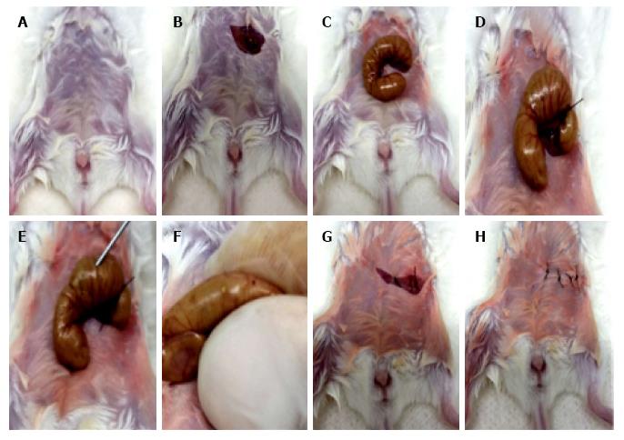
Figure 1 Main steps of the cecal ligation and puncture procedure.
A: Mice are anesthetized with i.p. administration of ketamine (100 mg/kg) and xylazine (12 mg/kg); B: After local asepsis, a cut is made in the skin and in the peritoneal membrane, providing access to the peritoneal cavity; C: The caecum is externalized through the incision and handled outside the peritoneal cavity; D: The caecum is ligated with suture thread right underneath the ileocaecal junction, but not so tightly that intestinal obstruction will ensue; E: The caecum is perforated with a needle, either on the proximal wall only [sublethal cecal ligation and puncture (CLP)] or completely transfixing (lethal CLP); F: The caecal contents are squeezed through the single (sublethal CLP) or double (lethal CLP) perforations; G: The caecum is repositioned inside the peritoneal cavity in its original location; H: The peritoneal membrane and the skin are sutured and the animals are undergo recovery from anesthesia protected from hypothermia and corneal damage or exposure to direct light. For sham-operated controls, steps D-G are omitted.
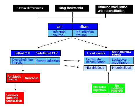
Figure 2 Modular distribution of multiple variables of interest in the cecal ligation and puncture surgical model.
The core parts of a cecal ligation and puncture (CLP) experiment (blue boxes) can be subdivided in modules that deal with trauma plus infection (CLP) or trauma without infection (sham). In both cases, events at the site of surgical injury (local events), i.e., the peritoneal cavity, and at a distant site (bone marrow events) can be observed in the same animals at any given time. Observation allows the experimenter to monitor progress of the host reaction (leukocyte accumulation in the peritoneal cavity; leukocyte mobilization from the bone marrow) as well as the dissemination of infectious pathogens (microbial load) at both sites. To this core, addition of preoperative modules (black boxes) involving a choice of different wild-type and mutant mouse strains (strain differences), a variety of prophylactic agents (drug treatments) and immunological interventions (immune modulation and reconstitution) considerably enriches the model in experimental possibilities. The issues of severity of infection (overwhelming infection vs severe infection, blue boxes at the left) and of the long-standing immune depression following recovery in antibiotic-rescued mice (red boxes at the left) are treated as separate modules of the trauma plus infection sector. Addition of postoperative modules (green boxes at the right) allows us to analyze the curative effect of mediators which restore mobilization of leukocytes from bone marrow into the peritoneal cavity, in mutants lacking 5-lipoxygenase (5-LO), or in wild-type mice preoperatively given inhibitors of the 5-LO pathway. In such a surgical model, every animal undergoes surgery, but the addition of genetical, pharmacological and immunological variables greatly enriches the model in its investigative power.
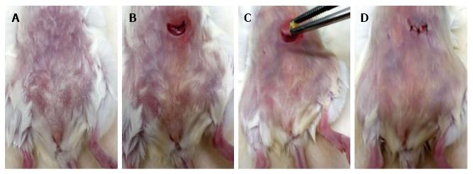
Figure 3 Main steps of the intraperitoneal modification of the egg white implant procedure.
A: Mice are anesthetized with i.p. administration of ketamine (100 mg/kg) and xylazine (12 mg/kg); B: After local asepsis, a cut is made in the skin and in the peritoneal membrane, providing access to the peritoneal cavity; C: A pellet of heat-coagulated egg white is placed through the opening in the peritoneal cavity; D: The peritoneal membrane and the skin are sutured and the animals are placed to recover from anesthesia with care to avoid hypothermia and corneal dryness or undue exposure to direct light. For sham-implanted controls, step C is omitted.
- Citation: Xavier-Elsas P, Ferreira RN, Gaspar-Elsas MIC. Surgical and immune reconstitution murine models in bone marrow research: Potential for exploring mechanisms in sepsis, trauma and allergy. World J Exp Med 2017; 7(3): 58-77
- URL: https://www.wjgnet.com/2220-315X/full/v7/i3/58.htm
- DOI: https://dx.doi.org/10.5493/wjem.v7.i3.58


