Copyright
©The Author(s) 2017.
World J Exp Med. Aug 20, 2017; 7(3): 58-77
Published online Aug 20, 2017. doi: 10.5493/wjem.v7.i3.58
Published online Aug 20, 2017. doi: 10.5493/wjem.v7.i3.58
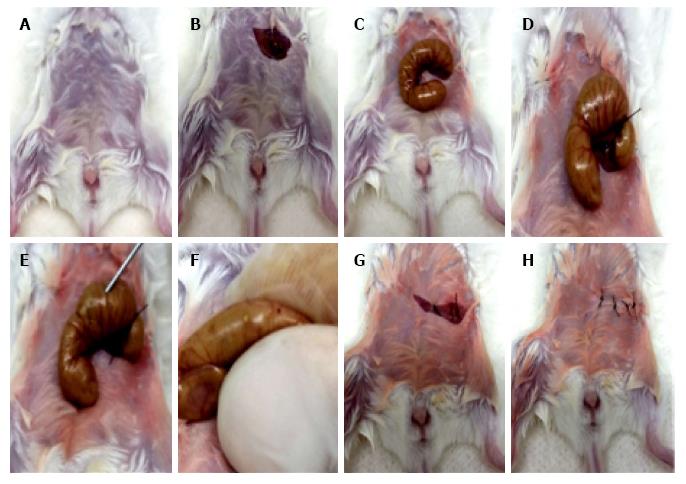
Figure 1 Main steps of the cecal ligation and puncture procedure.
A: Mice are anesthetized with i.p. administration of ketamine (100 mg/kg) and xylazine (12 mg/kg); B: After local asepsis, a cut is made in the skin and in the peritoneal membrane, providing access to the peritoneal cavity; C: The caecum is externalized through the incision and handled outside the peritoneal cavity; D: The caecum is ligated with suture thread right underneath the ileocaecal junction, but not so tightly that intestinal obstruction will ensue; E: The caecum is perforated with a needle, either on the proximal wall only [sublethal cecal ligation and puncture (CLP)] or completely transfixing (lethal CLP); F: The caecal contents are squeezed through the single (sublethal CLP) or double (lethal CLP) perforations; G: The caecum is repositioned inside the peritoneal cavity in its original location; H: The peritoneal membrane and the skin are sutured and the animals are undergo recovery from anesthesia protected from hypothermia and corneal damage or exposure to direct light. For sham-operated controls, steps D-G are omitted.
- Citation: Xavier-Elsas P, Ferreira RN, Gaspar-Elsas MIC. Surgical and immune reconstitution murine models in bone marrow research: Potential for exploring mechanisms in sepsis, trauma and allergy. World J Exp Med 2017; 7(3): 58-77
- URL: https://www.wjgnet.com/2220-315X/full/v7/i3/58.htm
- DOI: https://dx.doi.org/10.5493/wjem.v7.i3.58


