
















Copyright: ©Author(s) 2026.
World J Exp Med. Sep 20, 2026; 16(3): 125128
Published online Sep 20, 2026. doi: 10.5493/wjem.125128
Published online Sep 20, 2026. doi: 10.5493/wjem.125128
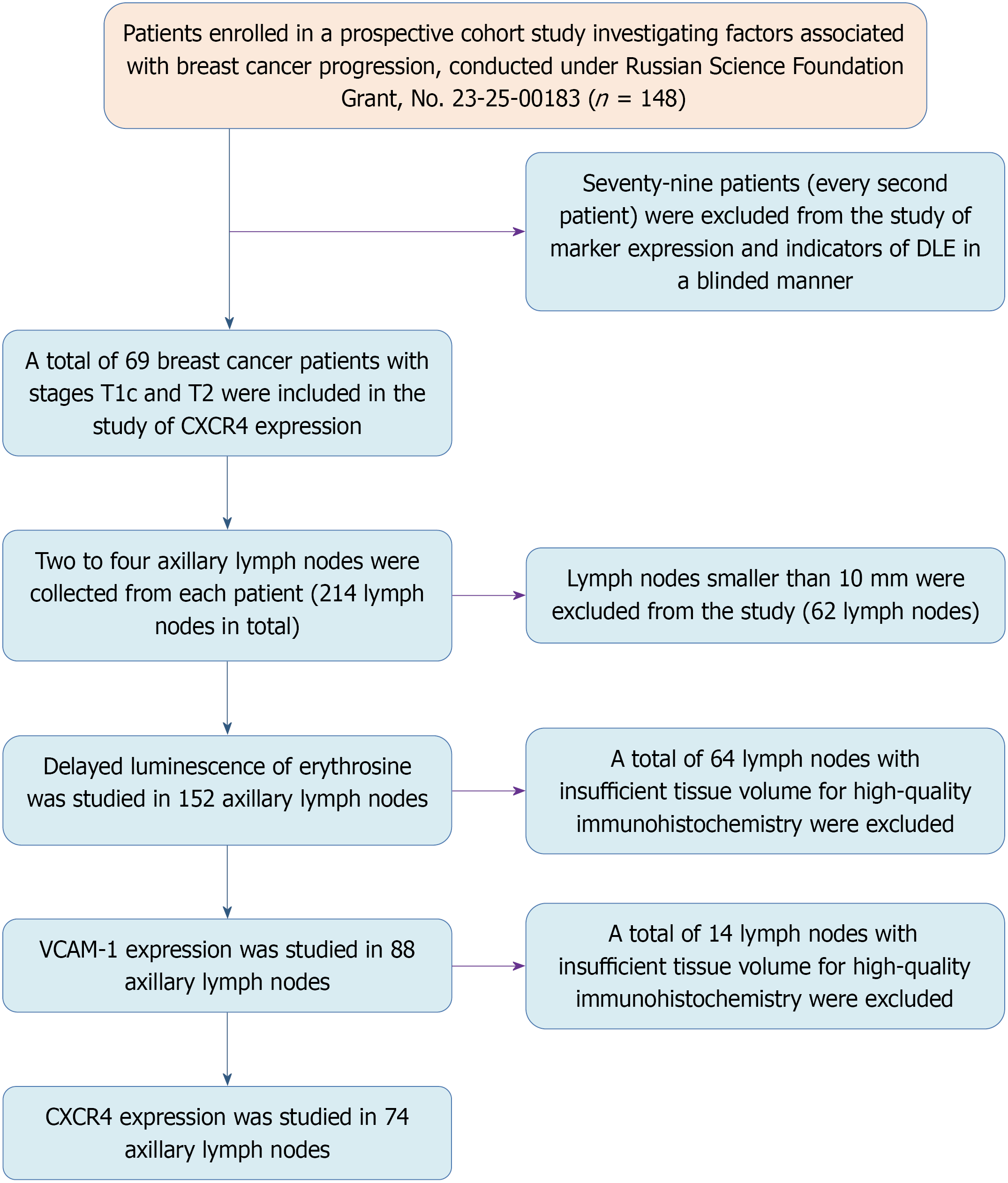
Figure 1 Study design flowchart.
DLE: Delayed luminescence of erythrosine; CXCR4: C-X-C motif chemokine receptor 4; VCAM-1: Vascular cell adhesion molecule-1.
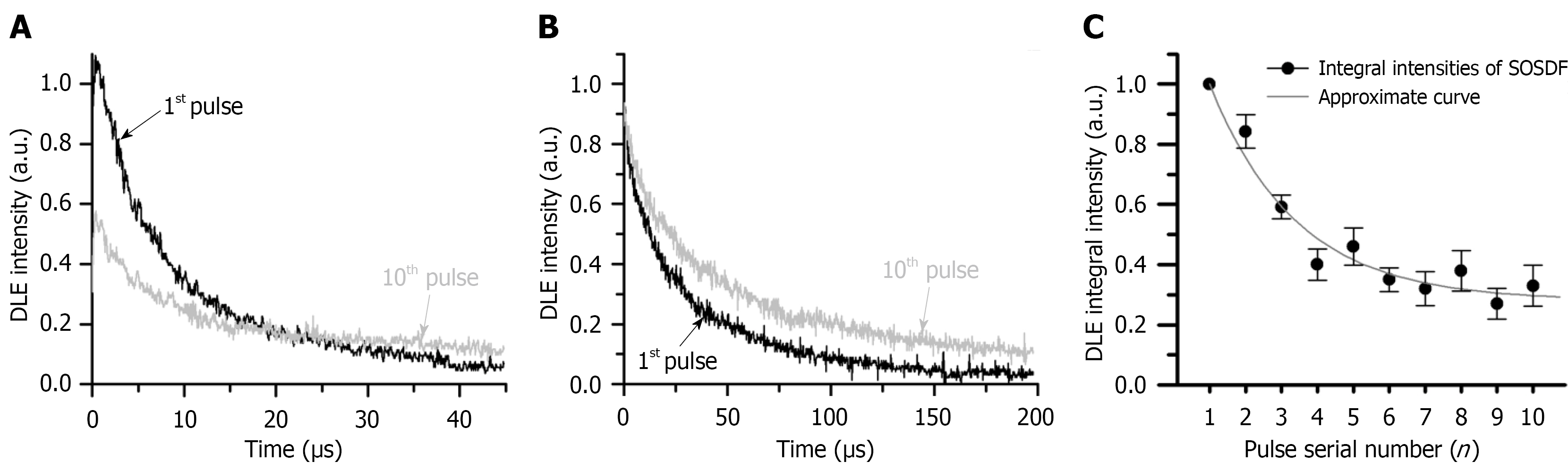
Figure 2 Kinetics of delayed fluorescence in a lymph node stained with erythrosine.
A: Kinetics of delayed fluorescence in a lymph node stained with erythrosine under multi-pulse excitation at a frequency of 10 Hz (curves for the first and tenth excitation pulses are shown); B: Kinetics of phosphorescence in a lymph node stained with erythrosine under multi-pulse excitation at a frequency of 10 Hz (curves for the first and tenth excitation pulses are shown); C: Change in the integral intensity of singlet oxygen-sensitized delayed fluorescence (SOSDF) of erythrosine in stained tissue during 10-pulse excitation (the integral intensity of SOSDF for the first pulse is taken as unity). SOSDF: Singlet oxygen-sensitized delayed fluorescence; DLE: Delayed luminescence of erythrosine.
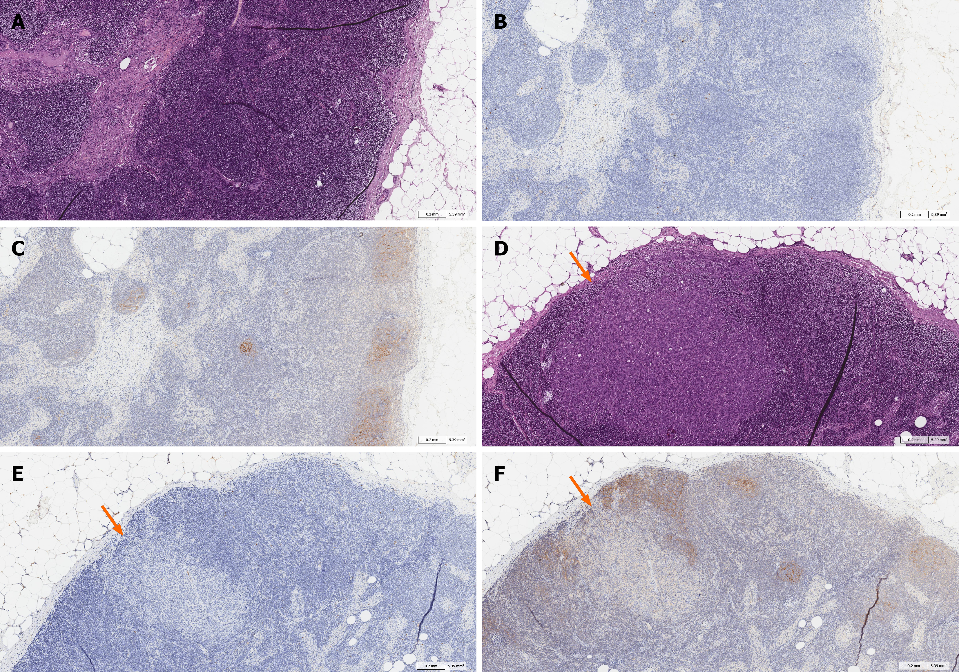
Figure 3 Examples of Vascular cell adhesion molecule-1 and C-X-C motif chemokine receptor 4 expression assessment in an axillary lymph node.
A: Assessment of the number of sinuses (2: > 15%) and lymphoid follicles (1: ≥ 5% and < 30% of the lymph node area) in the axillary lymph node; B: Assessment of vascular cell adhesion molecule-1 (VCAM-1) expression in the lymph node sinuses (0: No expression), lymph node follicles (0: No expression), lymph node cortex (0: No expression) and lymph node medulla (0: No expression); C: Assessment of C-X-C motif chemokine receptor 4 (CXCR4) expression in the lymph node sinuses (1: Weak expression), lymph node follicles (2: Moderate expression), lymph node cortex (1: Weak expression), and lymph node medulla (1: Weak expression); D: Breast cancer metastasis in the axillary lymph node (arrow); E: No VCAM-1 expression in tumor cells in the lymph node with metastasis; F: Weak CXCR4 expression in tumor cells in the lymph node with metastasis (A and D: Mayer’s hematoxylin and eosin staining; B and E: Staining with antibodies against VCAM-1; C and F: Staining with antibodies against CXCR4).
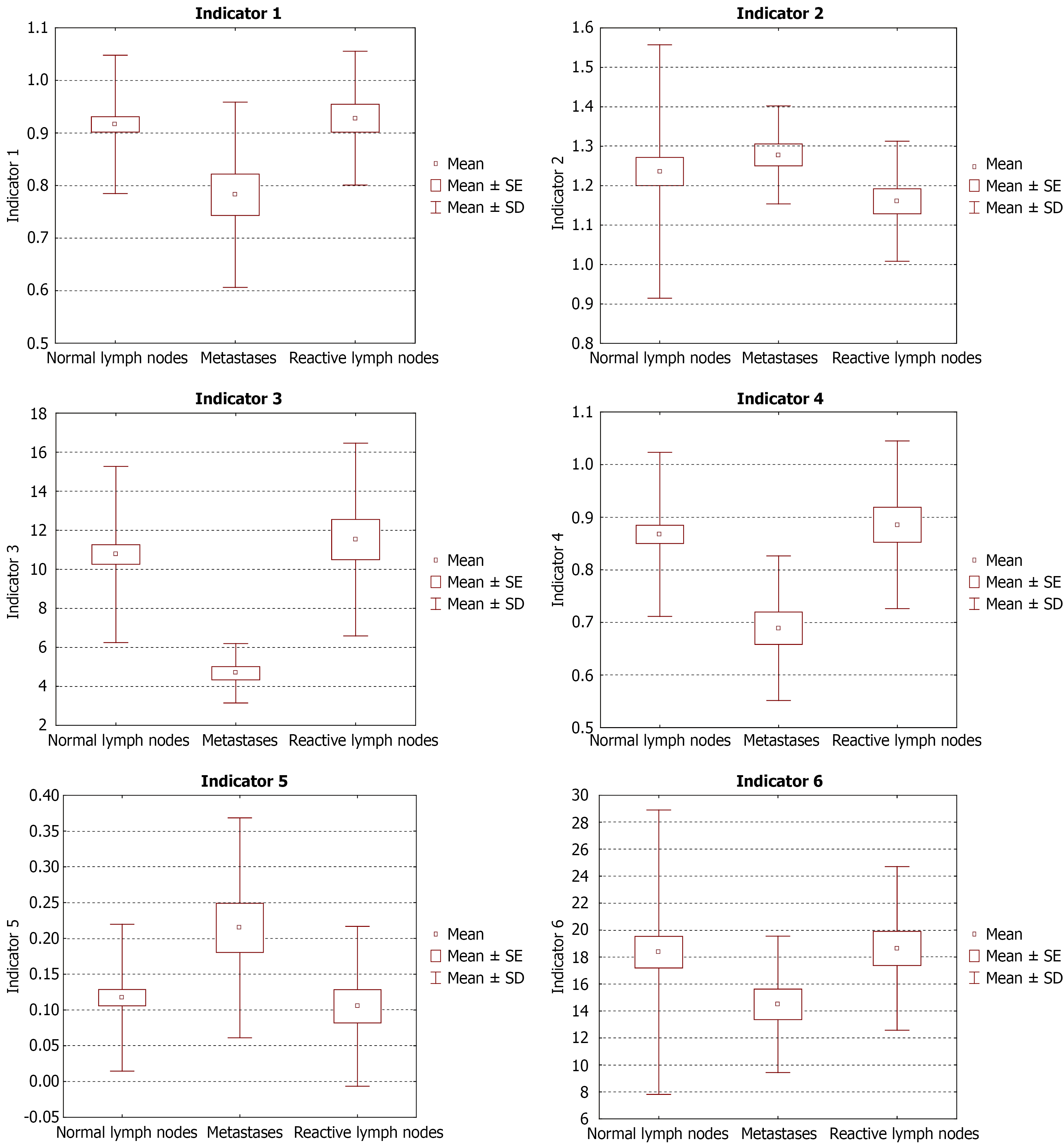
Figure 4 Values of the delayed luminescence of erythrosine depending on the nature of the changes in the axillary lymph nodes.
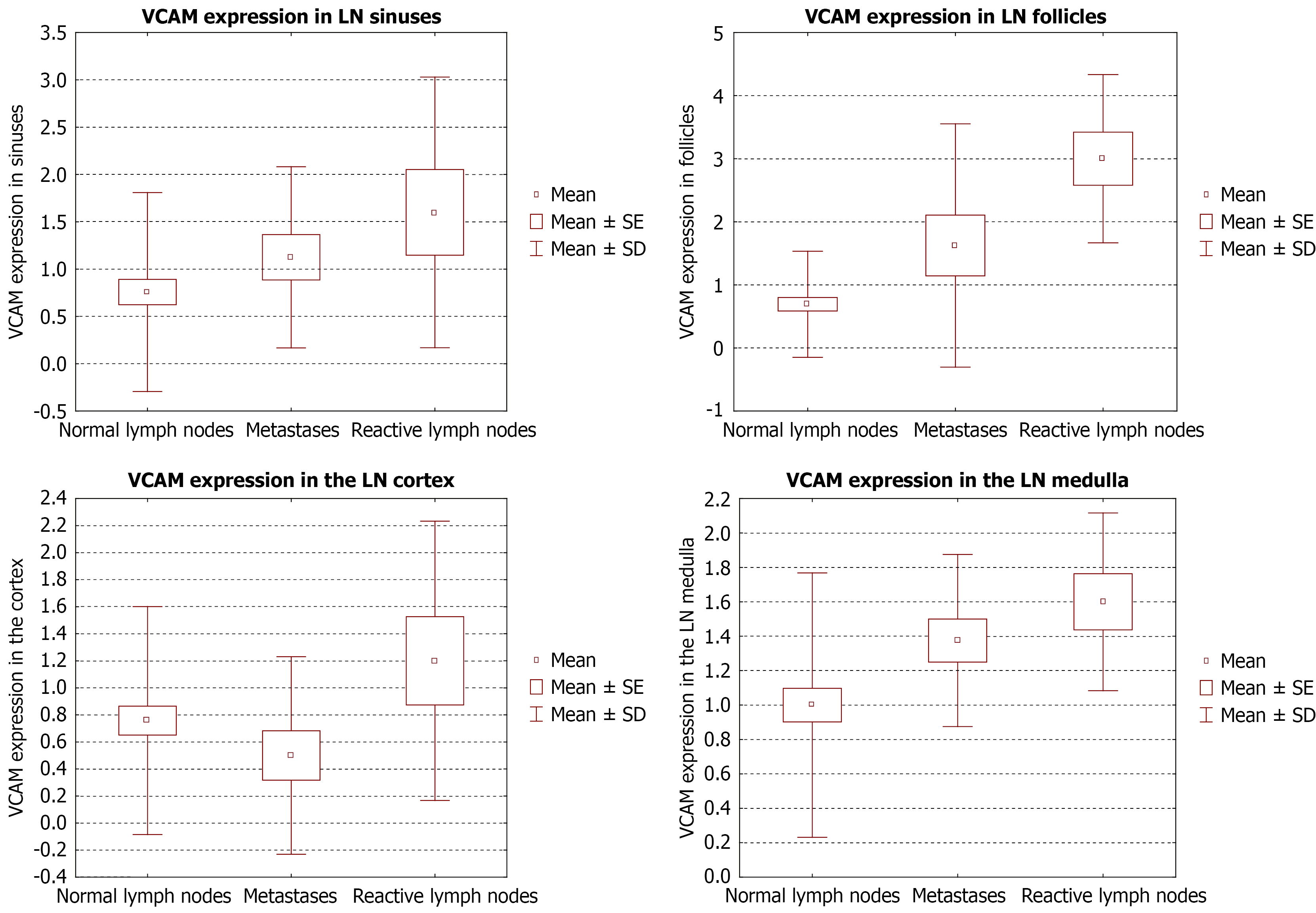
Figure 5 Vascular cell adhesion molecule-1 expression scores according to the nature of the changes in axillary lymph nodes.
VCAM-1: Vascular cell adhesion molecule-1; LN: Lymph node.
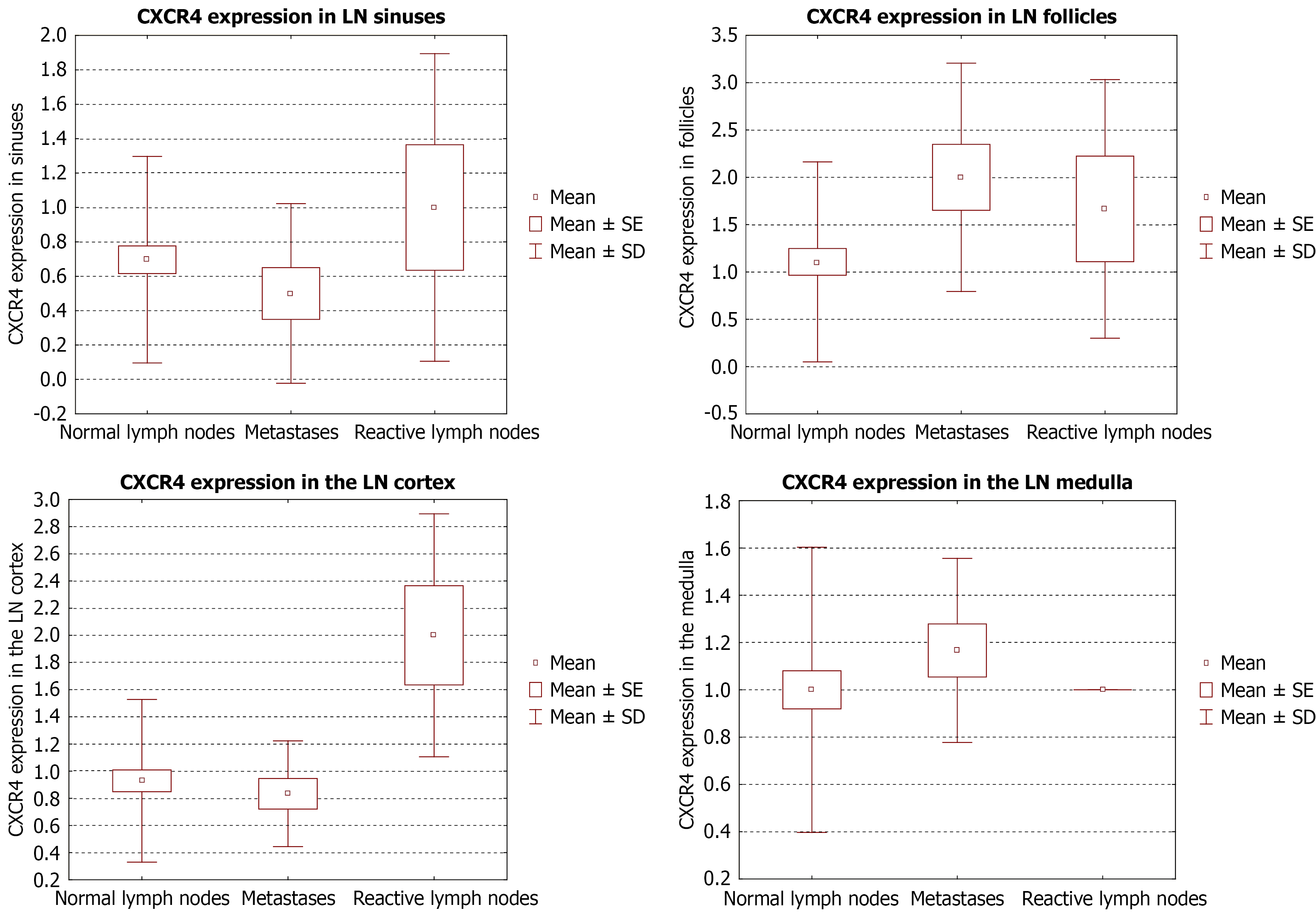
Figure 6 C-X-C motif chemokine receptor 4 expression scores depending on the nature of the changes in axillary lymph nodes.
CXCR4: C-X-C motif chemokine receptor 4; LN: Lymph node.
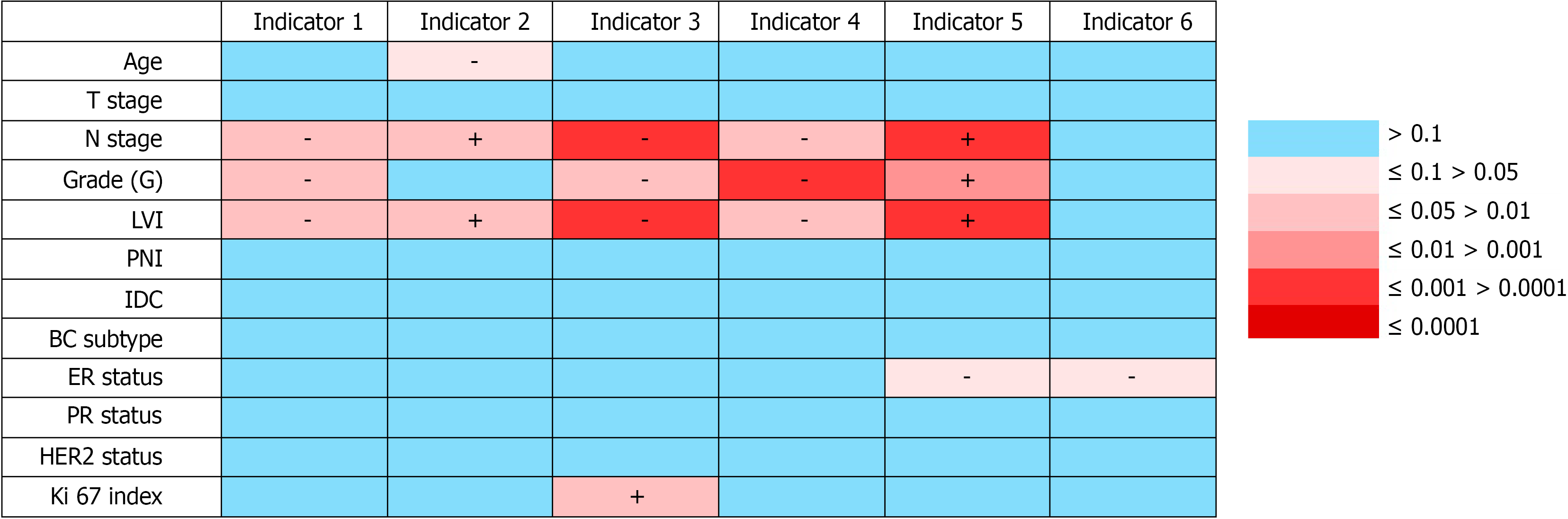
Figure 7 Heatmaps of correlations between indicators of delayed luminescence of erythrosine and clinical and pathological cha
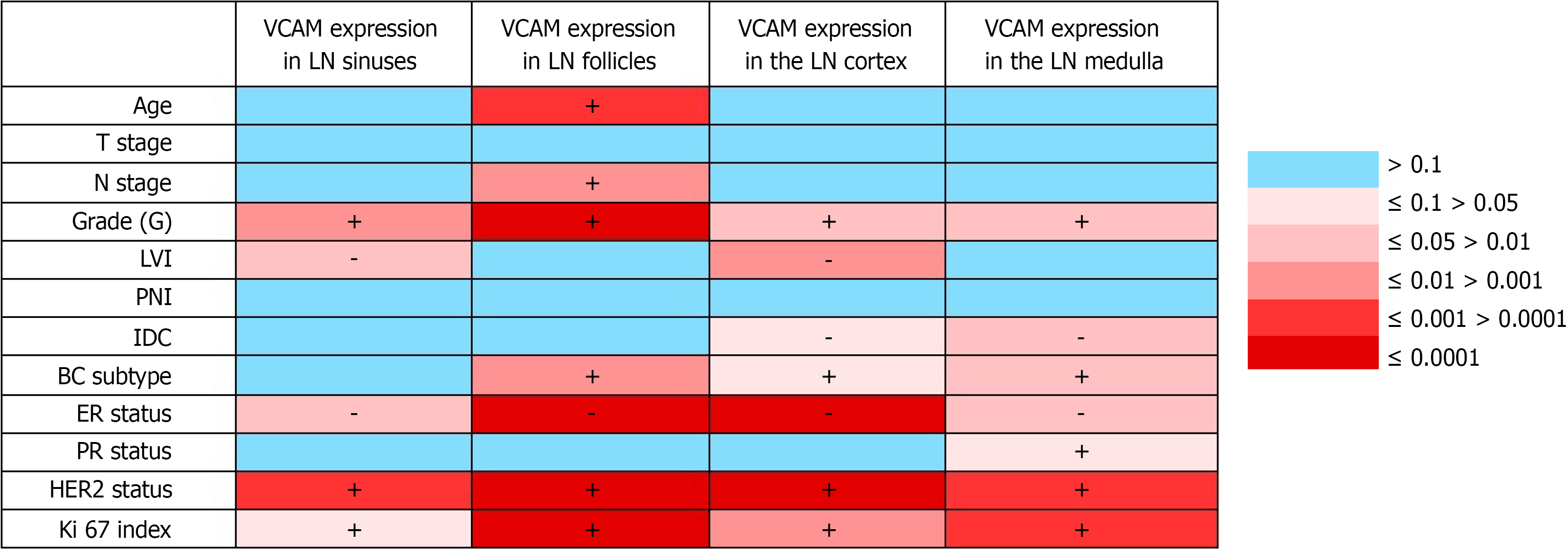
Figure 8 Heatmaps of correlations between vascular cell adhesion molecule-1 expression in axillary lymph nodes and clinical and pathological characteristics of patients with breast cancer.
VCAM: Vascular cell adhesion molecule; +: Positive correlations; -: Negative correlations; BC: Breast cancer; ER: Estrogen receptor; HER2: Human epidermal growth factor receptor 2; IDC: Intraductal component; LN: Lymph node; LVI: Lymphovascular invasion; PNI: Perineural invasion; PR: Progesterone receptor.
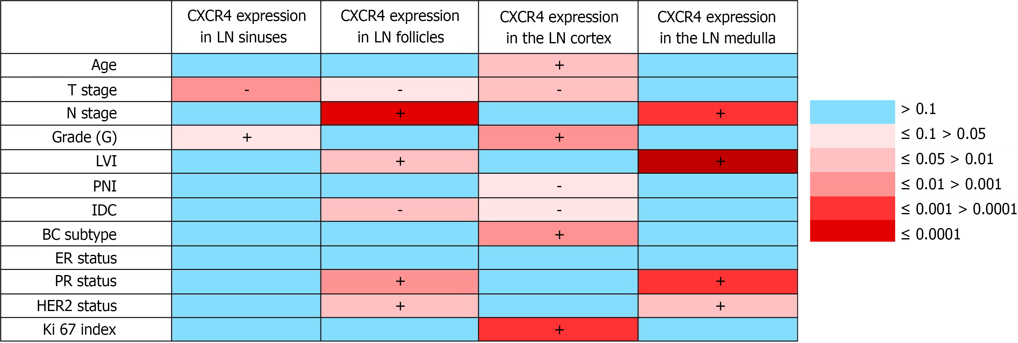
Figure 9 Heatmaps of correlations between C-X-C motif chemokine receptor 4 expression in axillary lymph nodes and clinical and pathological characteristics of patients with breast cancer.
+: Positive correlations; -: Negative correlations; CXCR4: C-X-C motif chemokine receptor 4; BC: Breast cancer; ER: Estrogen receptor; HER2: Human epidermal growth factor receptor 2; IDC: Intraductal component; LN: Lymph node; LVI: Lymphovascular invasion; PNI: Perineural invasion; PR: Progesterone receptor.
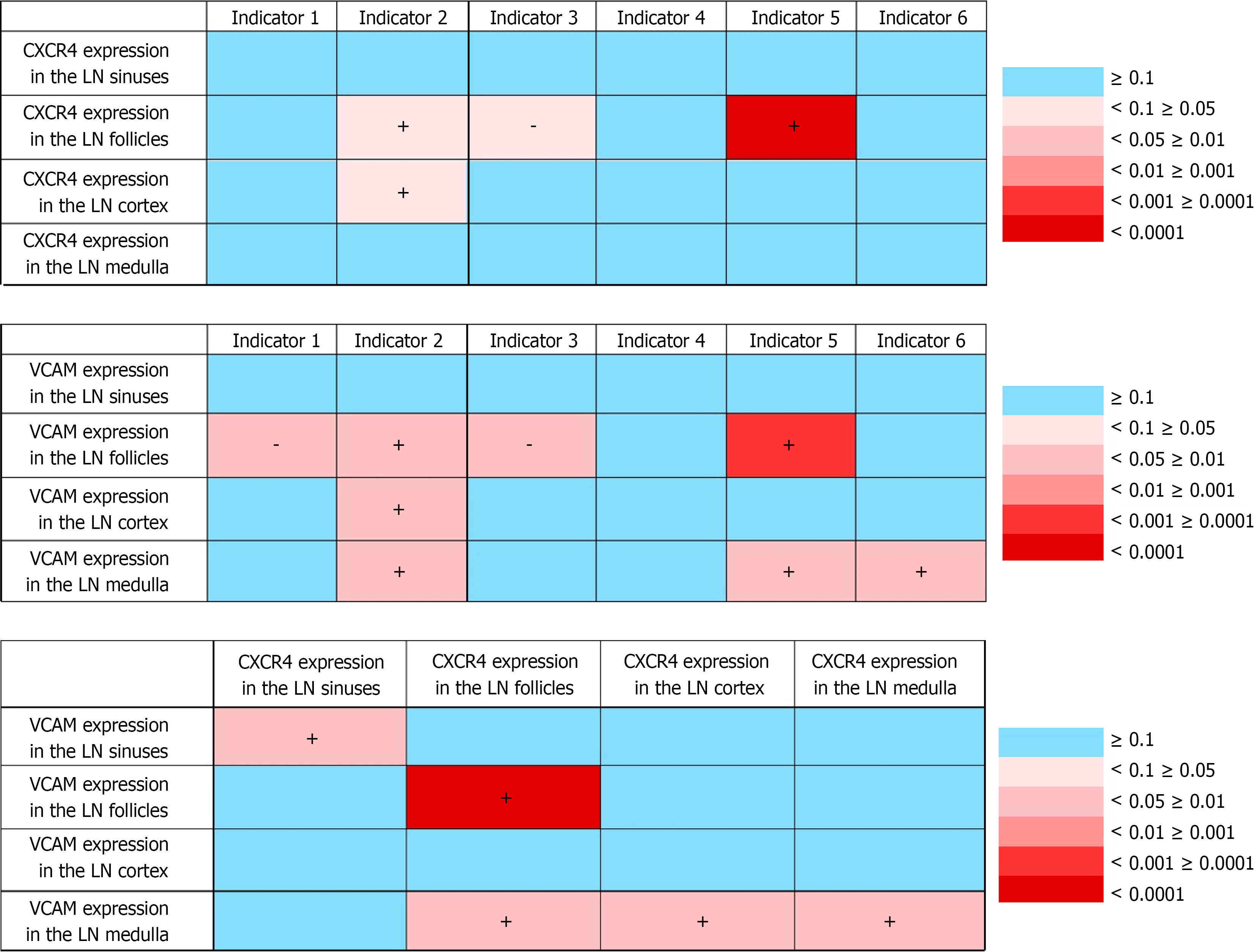
Figure 10 Correlations between the studied markers.
+: Positive correlations; -: Negative correlations; CXCR4: C-X-C motif chemokine receptor 4; LN: Lymph node; VCAM: Vascular cell adhesion molecule-1.
- Citation: Senchukova MA, Ishemgulov AT, Letuta SN, Saidler NV, Tagabilev DG. Changes in the metabolism of axillary lymph node cells as a predictor of regional metastasis in breast cancer. World J Exp Med 2026; 16(3): 125128
- URL: https://www.wjgnet.com/2220-315X/full/v16/i3/125128.htm
- DOI: https://dx.doi.org/10.5493/wjem.125128