Published online Mar 28, 2015. doi: 10.5412/wjsp.v5.i1.173
Peer-review started: September 10, 2014
First decision: September 28, 2014
Revised: January 15, 2015
Accepted: January 30, 2015
Article in press: February 2, 2015
Published online: March 28, 2015
Processing time: 204 Days and 12.8 Hours
We describe a novel technique for sentinel lymph node mapping and biopsy of a primary cutaneous malignant melanoma in the medial portion of the external auditory canal. The approach is illustrated through a case report and technical description of a procedure performed under general anesthesia on a 19-year-old female patient. Due to the hidden and sensitive location of the primary tumor in the medial external auditory canal, the lymphoscintigraphy injection had to be performed by the surgeon immediately prior to the resection of her cT2aN0M0 lesion. Final pathology revealed clear margins at the primary site resection and 2 intraparotid sentinel lymph nodes with microscopic foci of metastatic malignant melanoma, which led to further surgical management. A completion left parotidectomy and neck dissection yielded no additional metastatic disease in the fifty-five nodes that were evaluated. Using this technique, sentinel lymph node mapping and biopsy accurately predicted the highest risk lymph nodes for the primary lesion of the medial portion of the external auditory canal.
Core tip: We describe sentinel lymph node mapping and biopsy (SLNB) of a primary malignant melanoma in the external auditory canal. The usefulness of SLNB in this procedure allowed a focused surgical dissection to best assess regional lymph nodes and determine the extent of dissection needed to clear the disease. This novel technique is useful because it aids in establishing the single most important prognostic factor of a melanoma in the external auditory canal, regional lymph node status.
- Citation: Franco J, Hansen LA, Miyamoto RT, Tann M, Moore MG. Sentinel lymph node mapping for malignant melanoma of the external auditory canal. World J Surg Proced 2015; 5(1): 173-176
- URL: https://www.wjgnet.com/2219-2832/full/v5/i1/173.htm
- DOI: https://dx.doi.org/10.5412/wjsp.v5.i1.173
The incidence of malignant melanoma has surpassed that of other malignancies in recent years, at a rate of 3% per year[1]. In 2014, the American Cancer Society estimates that 76100 new cases of malignant melanoma would be diagnosed, resulting in 9710 deaths[2]. Of the malignant melanomas diagnosed, 18% are located in the head and neck and these are associated with a higher mortality rate when compared to melanomas of the trunk and extremities[1].
As treatment has evolved for melanoma, sentinel lymph node biopsy (SLNB) has been established as an integral aspect of both the surgical planning and treatment of this malignancy[3]. SLNB allows detection of minimal tumor burden and metastasis to the regional lymph nodes, an important prognostic factor in the American Joint Committee on Cancer staging system[4,5]. Other institutions including the National Comprehensive Cancer Network, the American Society of Clinical Oncology, and the Society of Surgical Oncology also recommend the routine use of SLNB. The status of regional lymph nodes portends the stratification of prognosis and assists with clinical trial determination and eligibility[6].
Melanoma of the external auditory canal is extremely rare, with fewer than 20 cases reported in the literature. The location of these lesions adds a level of difficulty to the standard surgical treatment, which is a wide local excision with margins of at least 1 cm[7]. In addition, technical aspects of radioisotope injection into the external auditory canal are more challenging because the ear canal skin is thin and requires additional instrumentation such as an otomicroscope for accessibility.
We present a case report of a patient with an intermediate stage (cT2aN0M0) cutaneous melanoma of the external auditory canal. As part of her management, a SLNB was performed. To our knowledge, this is the first description of this technique for a melanoma located in the bony portion of the external auditory canal.
Because this is a retrospective case report, per institutional protocol, no IRB approval was required. The patient has given consent to use her medical information for the case report. The medical records of the patient were accessed to obtain the clinical, radiographic and pathologic information.
The patient is a 19 years old, otherwise healthy female who presented with a complaint of a sore inside her left ear for approximately one year. The pigmented skin lesion was confined to the posteromedial aspect of the bony portion of the left external auditory canal. It did not involve the tympanic membrane nor did it involve the more lateral cartilaginous portion of the external auditory canal. There were no other skin lesions noted or any associated pathologically enlarged parotid or neck lymphadenopathy.
The patient was taken to the operating room where a biopsy was performed on the lesion, demonstrating a superficial malignant melanoma with a depth of invasion of 1.47 mm and no ulceration was observed. Appropriate staging revealed this to be a T2aN0M0 lesion. A combined positron emission tomography/computed tomography scan showed no evidence of regional lymphadenopathy or distant metastases; in addition, the bone underlying the lesion in the external auditory canal showed no erosion on preoperative imaging. After discussion with the patient, it was deemed appropriate for her to undergo resection of the lesion with a lateral temporal bone resection along with a SLNB. Due to the location of the lesion in the posteromedial aspect of the bony portion of the external auditory canal, it was most appropriate for the radioisotope injection to be performed using an otomicroscope under general anesthesia (we believe that an injection of local anesthetic to the ear canal skin would have distorted the lymphatic drainage of the area).
Using institutional protocol for handling of radioactive material, an aliquot of 1 mL of 1.5 mCi of Technetium labeled filtered sulfur colloid was obtained from the nuclear medicine department. A total of 0.15 mL was injected around the lesion (superior, inferior and lateral injections were used. No medial injection was performed due to the proximity to the tympanic membrane).
The patient was then prepped and draped in the normal sterile fashion and 30 min was allowed from the time of isotope injection to when the post-auricular incision was made to allow for diffusion through the lymphatics. Once the lateral temporal bone resection was completed, the hand held gamma probe was utilized to identify the most prominent sentinel lymph node which was located in the left parotid gland and had a 10 s count of 10381. The post-auricular incision was extended into the upper neck for access and this node was removed. A second lesion having a 10 s count of 4779 was also removed. The remaining parotid tail was noted to have a background higher than 10% of the original lymph node. As a result, the remaining parotid tail was removed and the associated wound background dropped to 389 on a 10 s count.
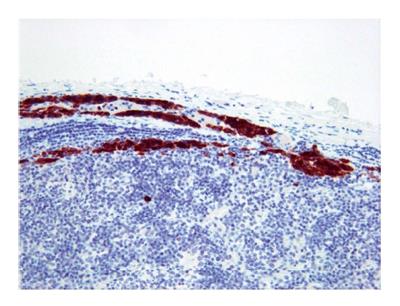
Surgical pathology of the primary lesion confirmed the 1.47 mm thick tumor and demonstrated that negative margins were obtained. Two of the seven lymph nodes removed as part of the sentinel lymph node mapping demonstrated microscopic foci of metastatic melanoma as demonstrated in Figure 1. As a result, it was recommended that the patient undergo a completion left parotidectomy and left modified radical neck dissection (MRND) with an abdominal fat graft for reconstruction. Final pathology from the completion parotidectomy and MRND showed no additional positive lymph nodes within the 55 that were removed in the completion procedure. The patient tolerated the procedure well and had no significant wound related complications or cranial nerve deficits. Due to the involvement of two intraparotid lymph nodes, the patient was referred to medical oncology. The patient subsequently underwent one year of interferon therapy and is now 20 mo post-op with no evidence of recurrent disease.
Malignant melanoma continues to claim lives and its incidence continues to increase at an alarming rate[1,2]. New techniques, such as SLNB, have developed to better serve these patients and reduce morbidity. The usefulness of SLNB in this procedure allowed a focused surgical dissection to best assess regional lymph nodes and determine the extent of dissection needed to clear the disease. This novel technique is useful because it aids in establishing the single most important prognostic factor of a melanoma in the external auditory canal, regional lymph node status[5,8-10]. Opinions on the use of SLNB for T1 lesions vary, while most authors agree on its use for T2 and T3 lesions. We demonstrate its utility in a smaller cancer located in the bony portion of the external auditory canal to accurately identify possible regional metastasis[5,6,11-13].
Studies have demonstrated that 15%-20% of patients with a primary head and neck melanoma will have lymph nodes in a regional nodal basin harboring occult metastases and that the sentinel lymph node accurately predicts the nodal basin status[14]. As such, SLNB plays an invaluable role in saving 80%-85% of patients with a primary head and neck malignant melanoma an unneeded lymph node dissection avoiding complications such as lymphedema, infection, hematoma, seroma, and nerve injury[12-15].
To date, a description of a SLNB of a malignant melanoma located in the bony portion of the external auditory canal does not exist in the literature. This paucity in literature creates uncertainty when dealing with this site-specific lesion. SLNB of the head and neck provides unique challenges related to the anatomy, technique, and interpretation of results[12]. The external auditory canal, due to its delicate and thin skin, as well as its poorly accessible location, is not easily amenable to standard sentinel lymph node mapping techniques. In our patient, the fact that both of the positive sentinel lymph nodes were the first two picked up at the time of the mapping indicates accuracy of the technique used. This is further supported by the fact that no additional positive nodes were obtained in the rest of the definitive lymphadenectomy.
We suggest that sentinel lymph node mapping for melanomas of the external auditory canal can be performed safely and successfully using our described technique.
Don-John Summerlin, DMD, MS for pathology photographs.
The 19-year-old female presented with a sore in the left ear for one year.
Pigmented skin lesion viewed with otoscopy, lesion confined to the posteromedial aspect of the bony portion of the left external auditory canal.
Malignant vs benign pigmented lesion.
All labs reviewed and within normal limits.
Positron emission tomography/computed tomography demonstrated no regional lymphadenopathy, no distant metastases, and no erosion of the bone underlying the lesion in the external auditory canal.
Initial biopsy demonstrated a superficial malignant melanoma with a depth of invasion of 1.47 mm and no ulceration. Two of the seven lymph nodes removed as part of the sentinel lymph node mapping demonstrated microscopic foci of metastatic melanoma.
Isotope injection with sentinel lymph node biopsy, lateral temporal bone resection, completion parotidectomy, and one year of interferon therapy.
To the best of the author’s knowledge, a description of a sentinel lymph node mapping and biopsy (SLNB) of a malignant melanoma located in the bony portion of the external auditory canal does not exist in the literature.
SLNB is a biopsy of the first lymph node or group of nodes draining an anatomic region. The sentinel lymph node is the first lymph node reached by metastasizing malignant cells.
This case presented unique challenges related to the anatomy, technique, and interpretation of results. The authors suggest that sentinel lymph node mapping for melanomas of the external auditory canal can be performed safely and successfully using our described technique.
The manuscript describes the application of lymph node mapping and biopsy in a case of primary malignant melanoma. It is well presented and easy to read.
P- Reviewer: Borrione P, Kimyai-Asadi A, Mazzocchi M
S- Editor: Gong XM L- Editor: A E- Editor: Liu SQ
| 1. | Saltman BE, Ganly I, Patel SG, Coit DG, Brady MS, Wong RJ, Boyle JO, Singh B, Shaha AR, Shah JP. Prognostic implication of sentinel lymph node biopsy in cutaneous head and neck melanoma. Head Neck. 2010;32:1686-1692. [RCA] [PubMed] [DOI] [Full Text] [Cited by in Crossref: 55] [Cited by in RCA: 58] [Article Influence: 4.1] [Reference Citation Analysis (0)] |
| 3. | Cappello ZJ, Augenstein AC, Potts KL, McMasters KM, Bumpous JM. Sentinel lymph node status is the most important prognostic factor in patients with melanoma of the scalp. Laryngoscope. 2013;123:1411-1415. [RCA] [PubMed] [DOI] [Full Text] [Cited by in Crossref: 19] [Cited by in RCA: 21] [Article Influence: 1.8] [Reference Citation Analysis (0)] |
| 4. | Egger ME, Callender GG, McMasters KM, Ross MI, Martin RC, Edwards MJ, Urist MM, Noyes RD, Sussman JJ, Reintgen DS. Diversity of stage III melanoma in the era of sentinel lymph node biopsy. Ann Surg Oncol. 2013;20:956-963. [RCA] [PubMed] [DOI] [Full Text] [Cited by in Crossref: 23] [Cited by in RCA: 23] [Article Influence: 1.8] [Reference Citation Analysis (0)] |
| 5. | Wong SL, Balch CM, Hurley P, Agarwala SS, Akhurst TJ, Cochran A, Cormier JN, Gorman M, Kim TY, McMasters KM. Sentinel lymph node biopsy for melanoma: American Society of Clinical Oncology and Society of Surgical Oncology joint clinical practice guideline. Ann Surg Oncol. 2012;19:3313-3324. [RCA] [PubMed] [DOI] [Full Text] [Cited by in Crossref: 89] [Cited by in RCA: 91] [Article Influence: 7.0] [Reference Citation Analysis (0)] |
| 6. | Gershenwald JE, Coit DG, Sondak VK, Thompson JF. The challenge of defining guidelines for sentinel lymph node biopsy in patients with thin primary cutaneous melanomas. Ann Surg Oncol. 2012;19:3301-3303. [RCA] [PubMed] [DOI] [Full Text] [Cited by in Crossref: 25] [Cited by in RCA: 25] [Article Influence: 2.1] [Reference Citation Analysis (0)] |
| 7. | Langman A, Yarington T, Patterson SD. Malignant melanoma of the external auditory canal. Otolaryngol Head Neck Surg. 1996;114:645-648. [RCA] [PubMed] [DOI] [Full Text] [Cited by in Crossref: 16] [Cited by in RCA: 16] [Article Influence: 0.6] [Reference Citation Analysis (0)] |
| 8. | Balch CM, Soong SJ, Gershenwald JE, Thompson JF, Reintgen DS, Cascinelli N, Urist M, McMasters KM, Ross MI, Kirkwood JM. Prognostic factors analysis of 17,600 melanoma patients: validation of the American Joint Committee on Cancer melanoma staging system. J Clin Oncol. 2001;19:3622-3634. [PubMed] |
| 9. | Mohebati A, Ganly I, Busam KJ, Coit D, Kraus DH, Shah JP, Patel SG. The role of sentinel lymph node biopsy in the management of head and neck desmoplastic melanoma. Ann Surg Oncol. 2012;19:4307-4313. [RCA] [PubMed] [DOI] [Full Text] [Cited by in Crossref: 35] [Cited by in RCA: 35] [Article Influence: 2.7] [Reference Citation Analysis (0)] |
| 10. | Morton DL, Cochran AJ, Thompson JF, Elashoff R, Essner R, Glass EC, Mozzillo N, Nieweg OE, Roses DF, Hoekstra HJ. Sentinel node biopsy for early-stage melanoma: accuracy and morbidity in MSLT-I, an international multicenter trial. Ann Surg. 2005;242:302-311; discussion 311-313. [PubMed] |
| 11. | Jones EL, Jones TS, Pearlman NW, Gao D, Stovall R, Gajdos C, Kounalakis N, Gonzalez R, Lewis KD, Robinson WA. Long-term follow-up and survival of patients following a recurrence of melanoma after a negative sentinel lymph node biopsy result. JAMA Surg. 2013;148:456-461. [RCA] [PubMed] [DOI] [Full Text] [Cited by in Crossref: 67] [Cited by in RCA: 81] [Article Influence: 6.8] [Reference Citation Analysis (0)] |
| 12. | Patel SG, Coit DG, Shaha AR, Brady MS, Boyle JO, Singh B, Shah JP, Kraus DH. Sentinel lymph node biopsy for cutaneous head and neck melanomas. Arch Otolaryngol Head Neck Surg. 2002;128:285-291. [PubMed] |
| 13. | van der Ploeg AP, van Akkooi AC, Verhoef C, Eggermont AM. Completion lymph node dissection after a positive sentinel node: no longer a must? Curr Opin Oncol. 2013;25:152-159. [RCA] [PubMed] [DOI] [Full Text] [Cited by in Crossref: 22] [Cited by in RCA: 22] [Article Influence: 1.8] [Reference Citation Analysis (0)] |
| 14. | Morton DL, Cochran AJ, Thompson JF. The rationale for sentinel-node biopsy in primary melanoma. Nat Clin Pract Oncol. 2008;5:510-511. [RCA] [PubMed] [DOI] [Full Text] [Full Text (PDF)] [Cited by in Crossref: 46] [Cited by in RCA: 45] [Article Influence: 2.6] [Reference Citation Analysis (0)] |
| 15. | Matter M, Lejeune FJ. The debate on sentinel node biopsy in melanoma: any clue? Melanoma Res. 2012;22:413-414. [RCA] [PubMed] [DOI] [Full Text] [Cited by in Crossref: 1] [Cited by in RCA: 1] [Article Influence: 0.1] [Reference Citation Analysis (0)] |


