Copyright
©2012 Baishideng.
World J Surg Proced. Jun 28, 2012; 2(3): 16-20
Published online Jun 28, 2012. doi: 10.5412/wjsp.v2.i3.16
Published online Jun 28, 2012. doi: 10.5412/wjsp.v2.i3.16
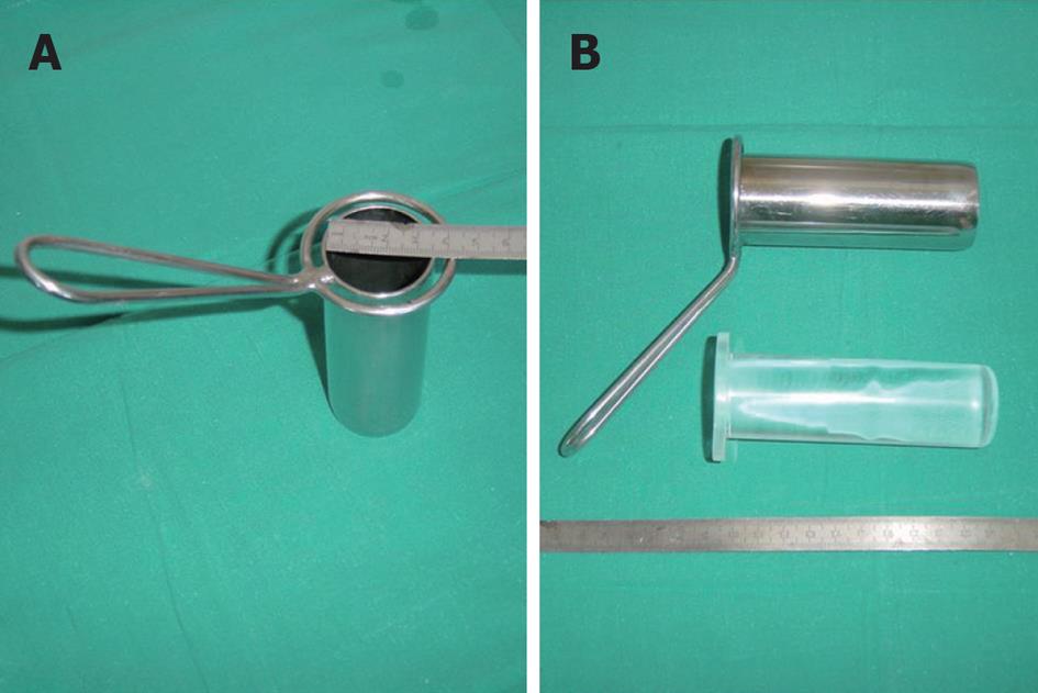
Figure 1 Cylinder used for transcylindrical cholecystectomy.
A: Dimensions of cylinders are 3.8 cm or 5.0 cm in diameter and 10.0 cm in length. The 5 cm cylinder is used in cases of known acute cholecystitis or when difficulties are anticipated or encountered at operation through the smaller cylinder; B: The cylinder has a transparent metacrilate plunger or embolus (like a piston) that, once the ensemble is introduced into the abdomen, allows visualization of the surgical field before unplugging.
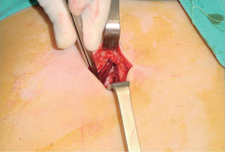
Figure 2 The cylinder is inserted through a transverse laparotomy incision over the right epigastrium, with dissection of the anterior rectus muscle fibers.
The incision uniformly measures 4.5 cm in length (or occasionally 7.5 cm if the 5 cm cylinder is used) throughout all layers of the abdominal wall.
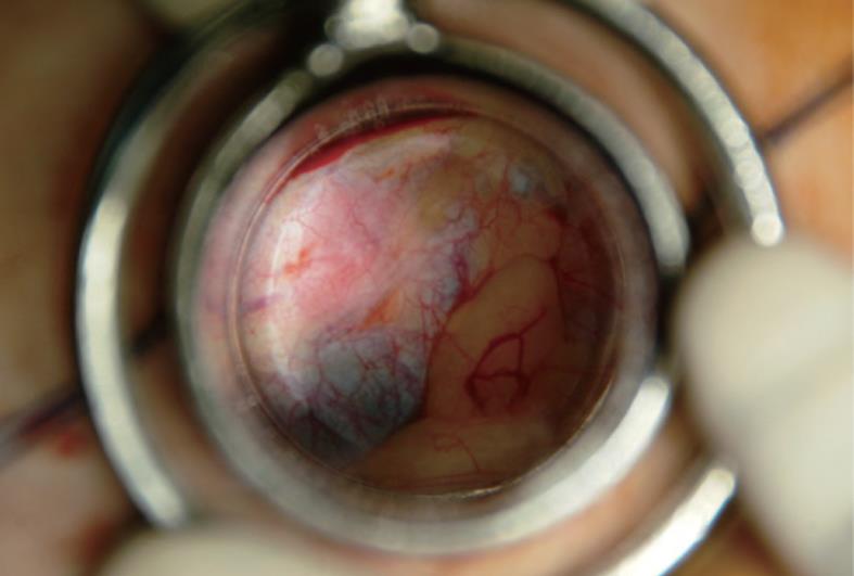
Figure 3 View through the metacrilate embolus of the cylinder.
The transparent plunger allows visualization of the surgical field before unplugging. The blunt shape of the plunger end, slightly protruding from the intra-abdominal side of the cylinder, facilitates the movement of the cylinder before reaching a working position at the hepatocystic triangle. Withdrawing the plunger does not cause any suction because it is slightly narrower than the lumen of the cylinder.
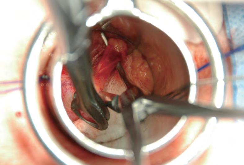
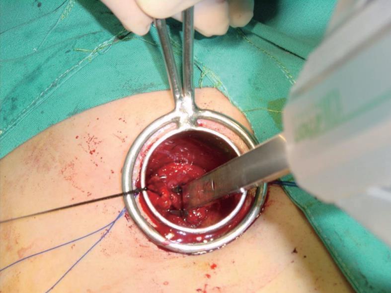
Figure 4 View through the cylinder during surgery.
The Hartmann Pouch is grasped with tissue forceps. Fat has been carefully dissected away using peanut gauze, as in conventional open surgery. The cystic duct, once clearly identified, is surrounded by using a right angle dissector. At this time, the surgeon and assistant must agree on the identity of the visible anatomic structures. The operation is carried out using reusable conventional open surgical instruments, the only exception being a disposable clip applier.
Figure 5 The cystic duct, about to be sectioned, has been ligated proximally and is being clipped distally.
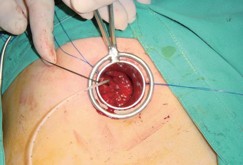
Figure 6 When cholangiography is required, a transcystic catheter is kept in place with a clip.
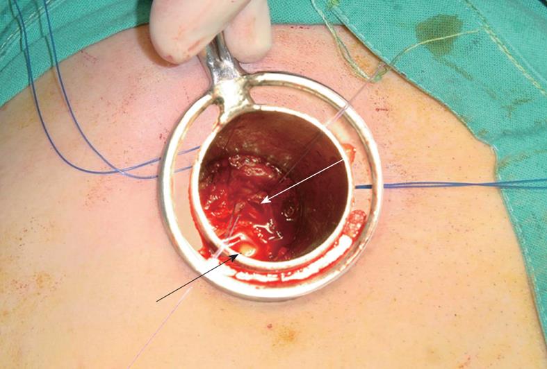
Figure 7 View through the 3.
8 cm cylinder. The common bile duct has been opened longitudinally between two tractor stitches (white arrow). A calculus has left the main biliary tree (black arrow). Randall’s Forceps cannot be used through the cylinder. The manoeuvres for exploration and stone removal are done using Fogarty’s Balloons, irrigation tubes or, occasionally, a choledochoscope.
- Citation: Grau-Talens EJ, Giner M. Technique of transcylindrical gas-free cholecystectomy. World J Surg Proced 2012; 2(3): 16-20
- URL: https://www.wjgnet.com/2219-2832/full/v2/i3/16.htm
- DOI: https://dx.doi.org/10.5412/wjsp.v2.i3.16


