Copyright
©The Author(s) 2016.
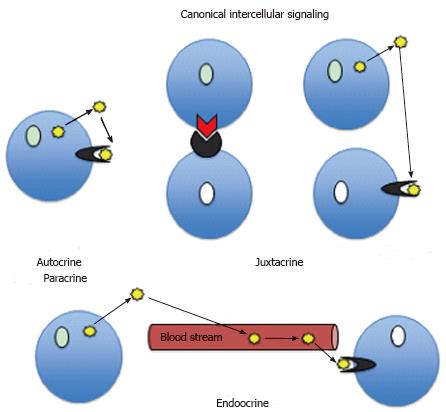
Figure 1 Types of canonical signaling.
In autocrine signaling, the cell regulates itself (autoregulates) through internally produced signaling molecules, which after release from the cell bind to cell’s own receptors. The examples are: Interleukin-1 produced by monocyte in response to external stimuli binds to its own receptor on the same monocyte; IL-2 released from activated T cell binds to its own receptor leading to self-stimulation. The juxtacrine signaling occurs between closely apposing cells when signaling molecule attached to one cell interacts with its receptor on adjacent cells or when signaling molecule excreted to the intercellular matrix of one cell binds to the receptor on neighboring cell. In juxtacrine signaling the signaling molecules do not diffuse freely between cells. The examples include cytokine signaling in immune system and Notch pathway signaling. In paracrine signaling, released signaling molecules such as, for example, cytokines or retinoic acid diffuse at short distances and act on the cells located in vicinity. In endocrine signaling, signaling molecules such as hormones or cytokines are transported through the circulation to the target cells.
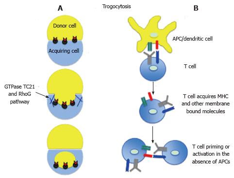
Figure 2 Trogocytosis/ membrane exchange/stripping.
A: During trogocytosis the recipient cell acquires membrane bound molecules from donor cell. The process of internalization of donor cell membrane is similar to phagocytosis and depends on actin ring contraction and small GTPases; B: During APC/T cell interaction the T cell may acquire MHC/peptide complexes and co-stimulatory molecules. Subsequently, such T cells can prime/activate naïve T cells in the absence of APCs, and/or by interacting with activated T cells lead to propagation of immune response[16,20]. APC: Antigen presenting cell; MHC: Major histocompatibility complex.
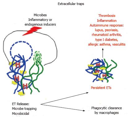
Figure 3 Extracellular traps formation and side effects.
Various external or internal inducers may lead to the breakage of nuclear or mitochondrial (or both) membranes and release of extracellular traps (ETs). The ETs contain network of nuclear/mitochondrial DNA (blue/green), antimicrobial compounds (yellow) such as LL37, myeoloperoxidase (lysosomal protein) and elastase (chymotrypsin-like protease), and deiminated (citrullinatied) histones (red H). Under normal circumstances the ETs are promptly removed by macrophages, however if the ETs persist they can lead to inflammatory and autoimmune response[31,32].
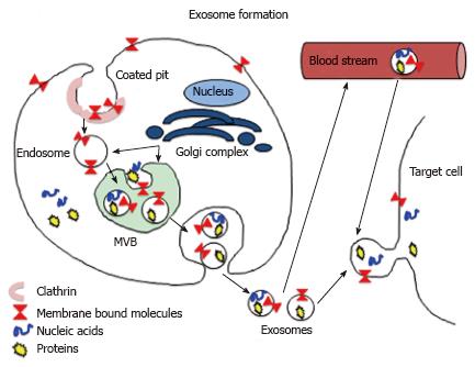
Figure 4 Exosome pathway.
Exosome forms through endocytosis, which starts from the invagination of clathrin coated domain of plasma membrane (coated pit) bearing the receptors and other membrane-bound molecules. After entering cell interior, the coated vesicle loses clathrin coat and becomes the endosome. Subsequently, after acquiring variety of other molecules from Golgi apparatus and cytoplasm, the endosome membrane undergoes inward budding resulting in the formation of multivesiular multivesicular body (MVB) containing exososmes. Ultimately, the MVB fuses with plasma membrane and in the process of exocytosis releases exosomes outside the cell. The exososmes either fuse with the membrane of neighboring target cell or enter blood stream to be transported to distant targets. In the alternative outcome (not shown here), which serves as the pathway degradation pathway, the MVB fuses with lysosomes, which degrade its content[67,69].
- Citation: Kloc M, Kubiak JZ, Li XC, Ghobrial RM. Noncanonical intercellular communication in immune response. World J Immunol 2016; 6(1): 67-74
- URL: https://www.wjgnet.com/2219-2824/full/v6/i1/67.htm
- DOI: https://dx.doi.org/10.5411/wji.v6.i1.67


