Copyright
©The Author(s) 2016.
World J Clin Pediatr. May 8, 2016; 5(2): 223-227
Published online May 8, 2016. doi: 10.5409/wjcp.v5.i2.223
Published online May 8, 2016. doi: 10.5409/wjcp.v5.i2.223
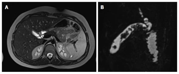
Figure 1 T2-weighted magnetic resonance imaging images confirming the irregular and diffuse thickening of gallbladder walls, more prominent in the body and in the fundus, suggestive of diffuse adenomyomatosis of the gallbladder (A and B).
Nodular images with intraluminal protrusion were localized in the fundus and in the body of the gallbladder.
Figure 2 Intraoperative picture.
A: Gallbladder specimen of 7 cm × 2 cm; B: Pathological examination showing the invagination of the gland (asterisks) into the muscular layer of the gallbladder, with the presence of glandular elements in the outer layers of the organ (hematoxilin-eosin, original magnification 4 ×); C: Heterotopic pancreatic glands (asterisks) in the context of the mucosal layer of the gallbladder (hematoxilin-eosin, original magnification 20 ×).
- Citation: Parolini F, Indolfi G, Magne MG, Salemme M, Cheli M, Boroni G, Alberti D. Adenomyomatosis of the gallbladder in childhood: A systematic review of the literature and an additional case report. World J Clin Pediatr 2016; 5(2): 223-227
- URL: https://www.wjgnet.com/2219-2808/full/v5/i2/223.htm
- DOI: https://dx.doi.org/10.5409/wjcp.v5.i2.223


