Copyright
©The Author(s) 2025.
World J Clin Pediatr. Sep 9, 2025; 14(3): 104096
Published online Sep 9, 2025. doi: 10.5409/wjcp.v14.i3.104096
Published online Sep 9, 2025. doi: 10.5409/wjcp.v14.i3.104096
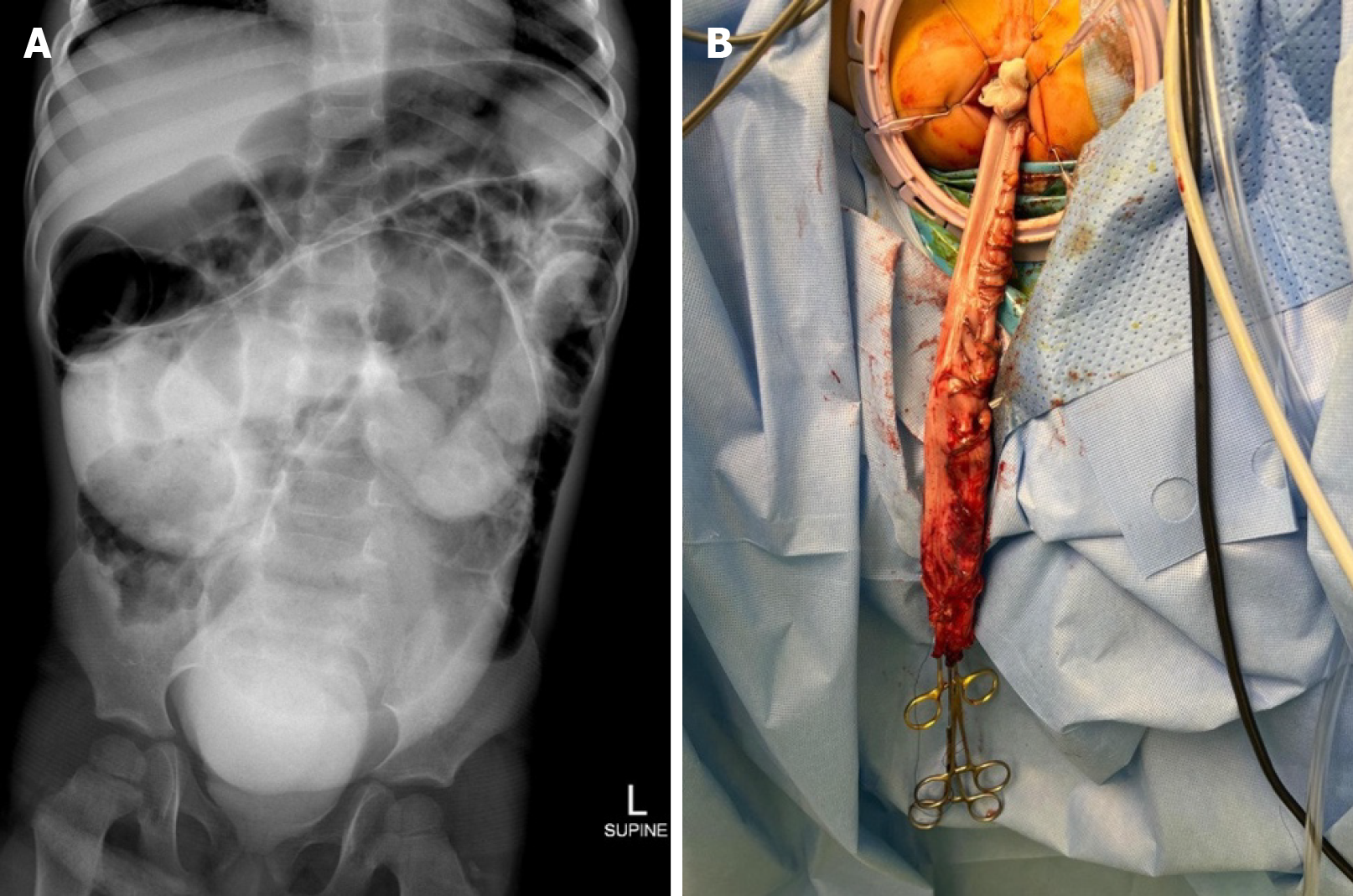
Figure 1 Radiological and surgical images from Case 1.
A: Rectally administered contrast revealed opacity and a markedly distended colonic structure in the central abdomen and pelvis; B: The distal sigmoid colon was pulled down from the anal verge as a part of a laparoscopic pull-through procedure.
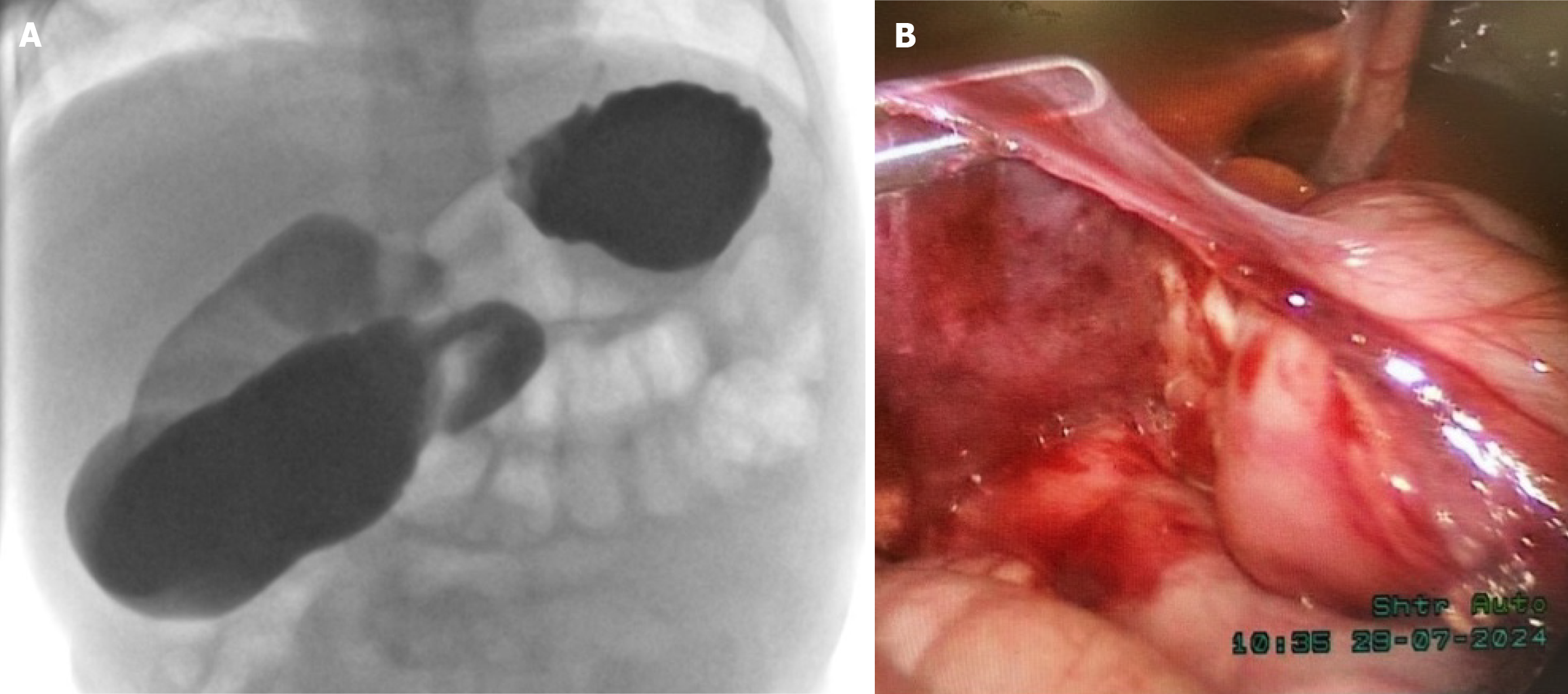
Figure 2 Radiological and surgical images from Case 2.
A: Barium swallow showed contrast passing from the stomach to duodenum with significant dilation to the duodenal jejunal junction, which was located in the midline and did not cross to the left side; B: A laparoscopic Ladd’s procedure was performed. The duodenum was isolated, and the Ladd’s bands were resected.
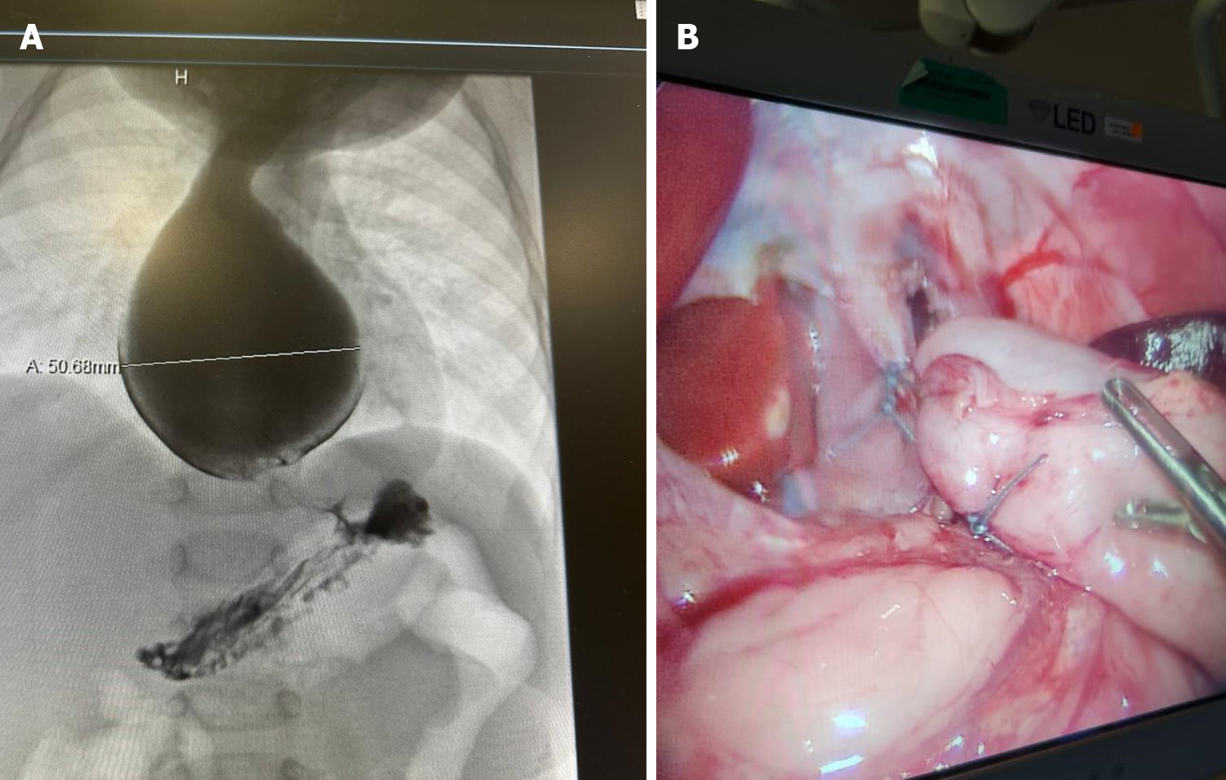
Figure 3 Radiological and surgical images for Case 3.
A: An upper gastrointestinal contrast study showed significant dilation of the esophagus with a pooling of contrast and holdup at the gastroesophageal junction; B: Laparoscopic Heller myotomy was performed.
- Citation: Shah R, Belsha D, Thomas A, Alsweed A. High suspicion unveils Hidden pathology of pediatric gastrointestinal surgical cases misidentified as medical: Three case reports. World J Clin Pediatr 2025; 14(3): 104096
- URL: https://www.wjgnet.com/2219-2808/full/v14/i3/104096.htm
- DOI: https://dx.doi.org/10.5409/wjcp.v14.i3.104096


