Copyright
©The Author(s) 2025.
World J Clin Pediatr. Sep 9, 2025; 14(3): 104096
Published online Sep 9, 2025. doi: 10.5409/wjcp.v14.i3.104096
Published online Sep 9, 2025. doi: 10.5409/wjcp.v14.i3.104096
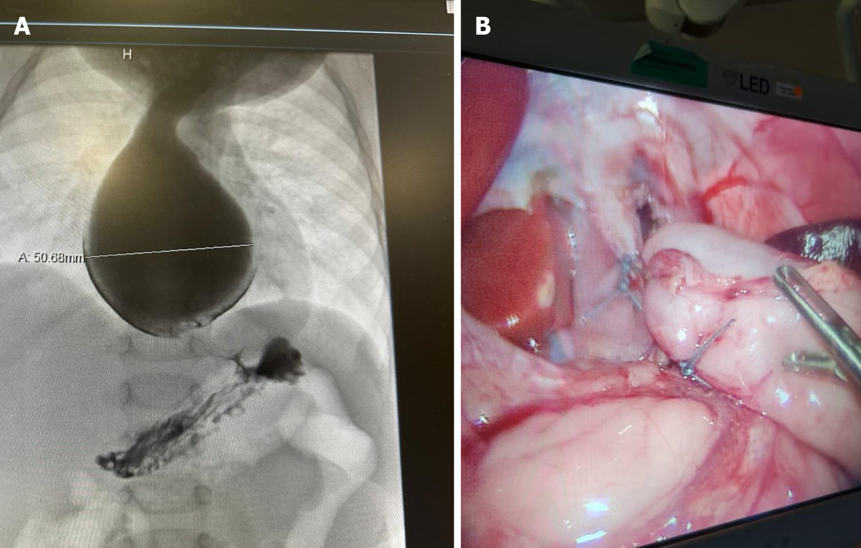
Figure 3 Radiological and surgical images for Case 3.
A: An upper gastrointestinal contrast study showed significant dilation of the esophagus with a pooling of contrast and holdup at the gastroesophageal junction; B: Laparoscopic Heller myotomy was performed.
- Citation: Shah R, Belsha D, Thomas A, Alsweed A. High suspicion unveils Hidden pathology of pediatric gastrointestinal surgical cases misidentified as medical: Three case reports. World J Clin Pediatr 2025; 14(3): 104096
- URL: https://www.wjgnet.com/2219-2808/full/v14/i3/104096.htm
- DOI: https://dx.doi.org/10.5409/wjcp.v14.i3.104096


