Copyright
©The Author(s) 2019.
World J Stomatol. Feb 20, 2019; 7(2): 20-27
Published online Feb 20, 2019. doi: 10.5321/wjs.v7.i2.20
Published online Feb 20, 2019. doi: 10.5321/wjs.v7.i2.20
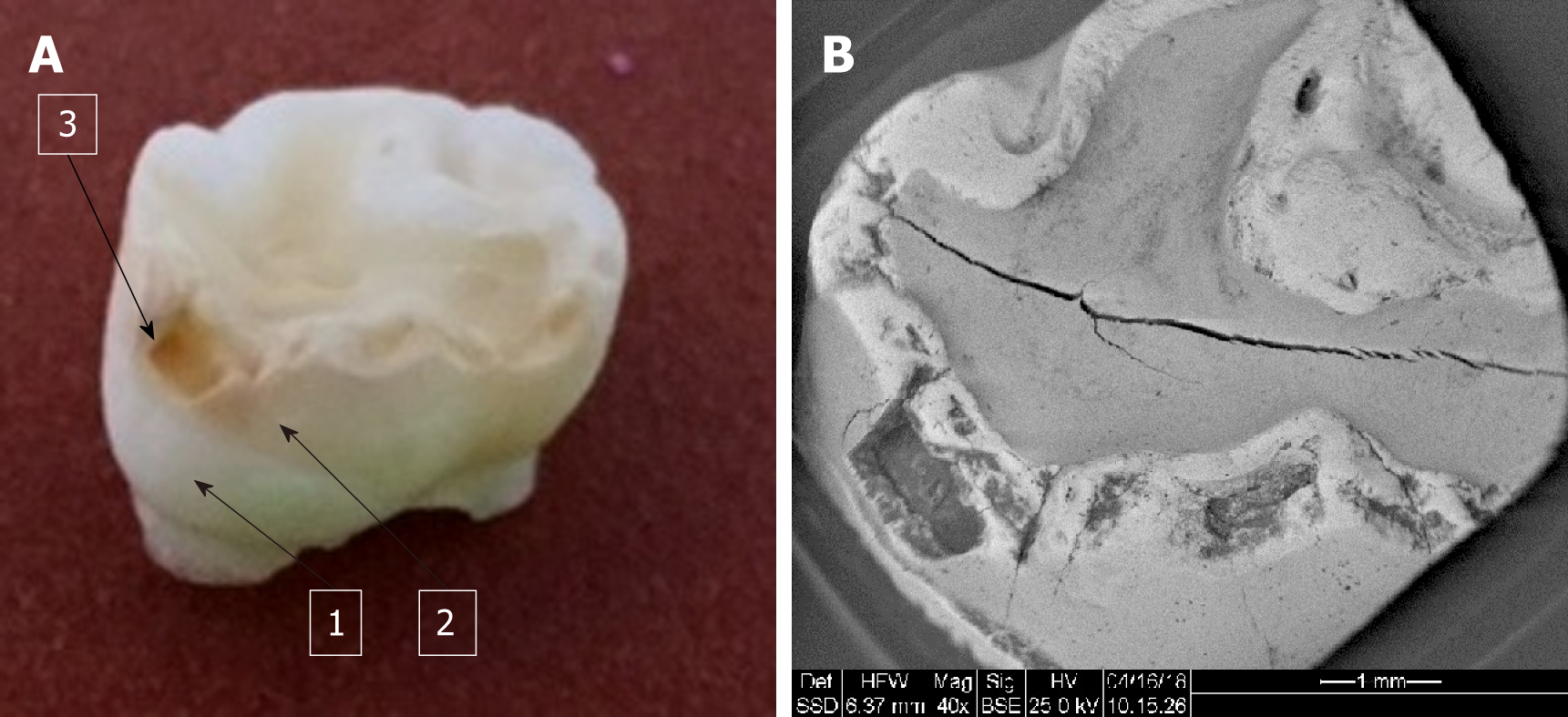
Figure 1 Clinical and SEM pictures of hypomineralized first primary molar.
A: Clinical picture of hypomineralized first primary molar. 1: Normal enamel; 2: Brown discolored enamel; 3: Breakdown of enamel; B: SEM picture of hypomineralized first primary molar.
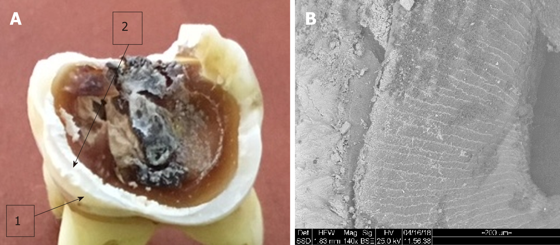
Figure 2 Clinical image of the first permanent molar affected by MH and the SEM image of the analyzed enamel.
A: Clinical picture. 1: Normal enamel; 2: Hypomineralized enamel border; B: SEM picture.
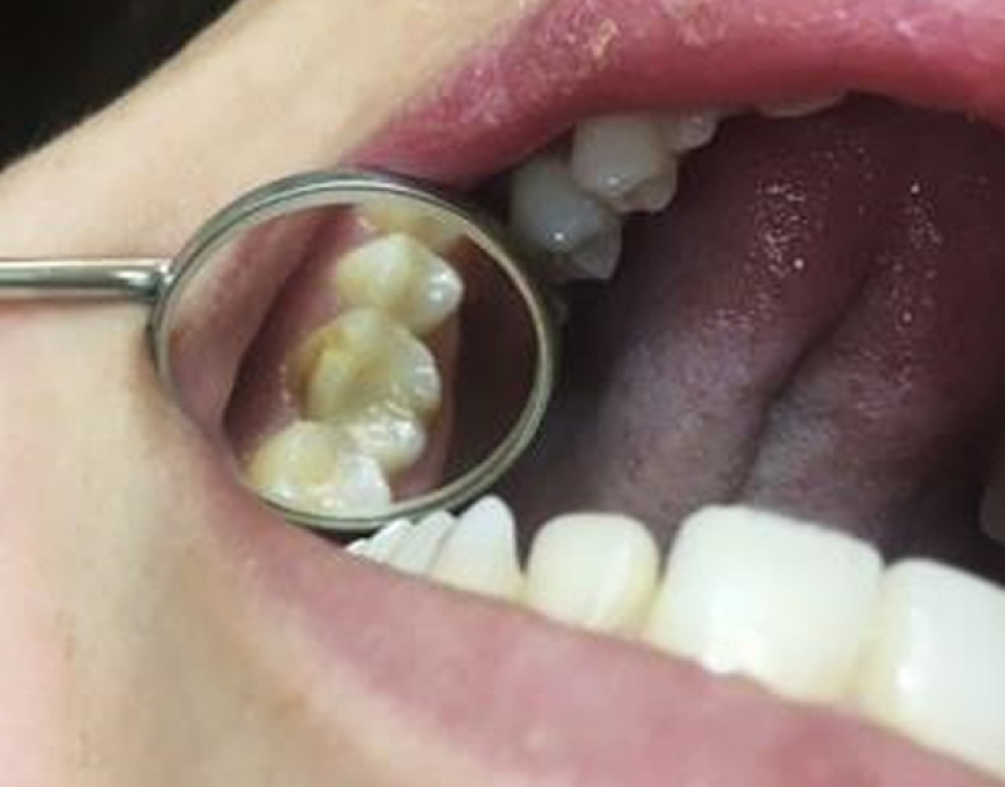
Figure 3 Restoration of disto-buccal cusp of upper permanent M1 affected by MIH with Glass-ionomer cement (EQUIA forte by GC Japan).
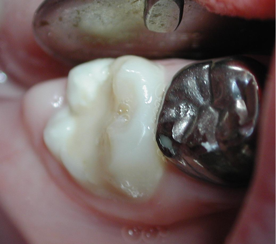
Figure 4 Bucco-occlusal restoration of permanent lower first molar affected by MIH with Glass-ionomer cement (EQUIA forte by GC Japan).
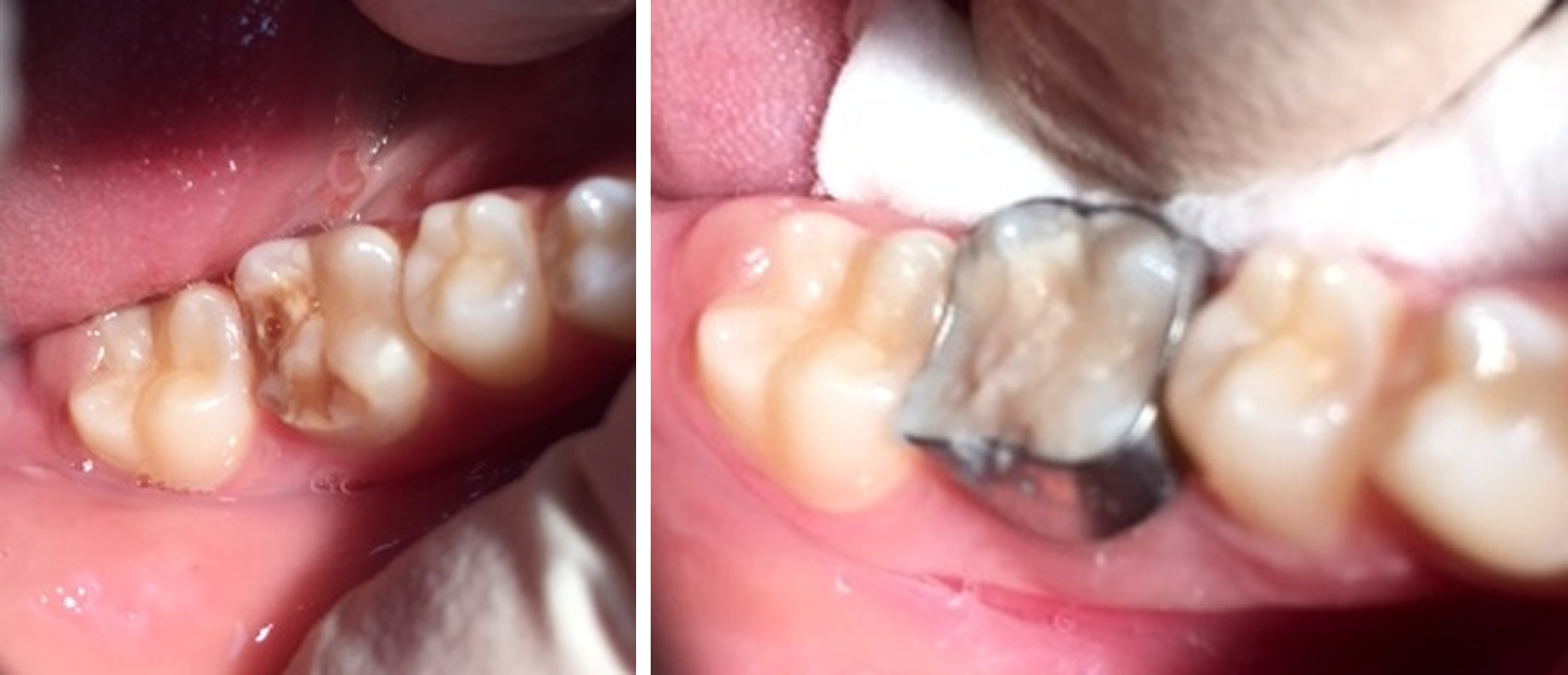
Figure 5 Breakdown of enamel of a first permanent molar due to MIH.
An orthodontic ring was cemented with Fuji 1 (Glass-ionomer cement) and the lesion filled with Glass-ionomer cement (EQUIA forte by GC Japan).
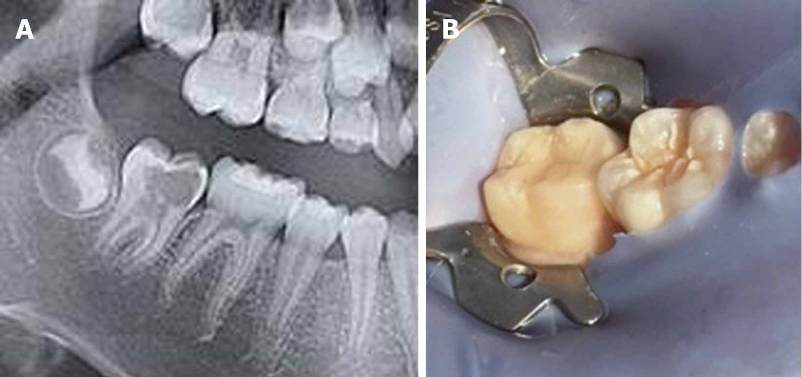
Figure 6 Pre-eruptive intracoronal lesions of unerrupted second permanent molar (A) and restoration of the lesion with Glass-ionomer cement (EQUIA forte by GC Japan).
A: SEM picture; B: Clinical picture.
- Citation: Zilberman U, Hassan J, Leiboviz-Haviv S. Molar incisor hypomineralization and pre-eruptive intracoronal lesions in dentistry-diagnosis and treatment planning. World J Stomatol 2019; 7(2): 20-27
- URL: https://www.wjgnet.com/2218-6263/full/v7/i2/20.htm
- DOI: https://dx.doi.org/10.5321/wjs.v7.i2.20


