Copyright
©The Author(s) 2015.
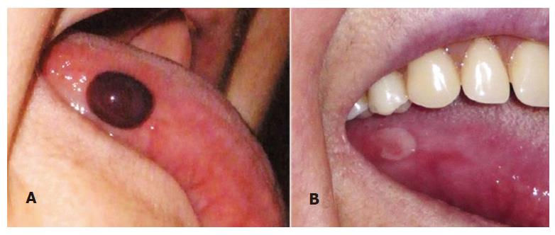
Figure 1 Clinical presentation of the disease.
A: Blister on the right lateral border of the tongue; B: Superficial ulcer after rupture of the hemorrhagic bulla (4 d of evolution).
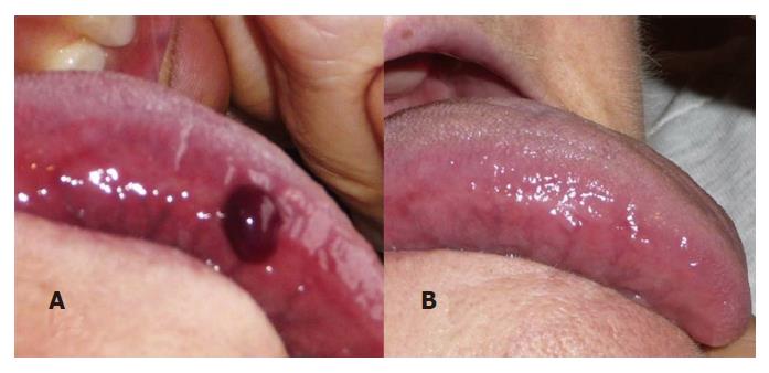
Figure 2 Presentation and resolution of a clinical case.
Blister with blood content (angina bullosa hemorrhagica) on the border of the tonge (A) and full clinical resolution after 14 d of evolution (B).
- Citation: Alberdi-Navarro J, Gainza-Cirauqui ML, Prieto-Elías M, Aguirre-Urizar JM. Angina bullosa hemorrhagica an enigmatic oral disease. World J Stomatol 2015; 4(1): 1-7
- URL: https://www.wjgnet.com/2218-6263/full/v4/i1/1.htm
- DOI: https://dx.doi.org/10.5321/wjs.v4.i1.1


