Copyright
©2014 Baishideng Publishing Group Inc.
World J Stomatol. Aug 20, 2014; 3(3): 25-29
Published online Aug 20, 2014. doi: 10.5321/wjs.v3.i3.25
Published online Aug 20, 2014. doi: 10.5321/wjs.v3.i3.25
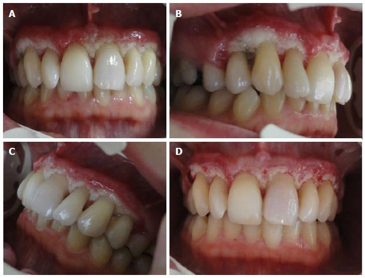
Figure 1 Intraoral photographs of the patient.
A: Frontal; B: Right; C: Left; D: Perforations on cortical bone prepared for clot formation.
Figure 2 Three months later intraoral photos of partially healed gums.
A: Frontal; B: Right; C: Left.
Figure 3 After a year all denuded areas were completely covered with gingivae and acceptable aesthetic and function have been formed.
A: Frontal; B: Right; C: Left.
Figure 4 Histopathologic comparing of gingiva samples.
A: Healthy gingiva; mild inflammatory cell infiltration (PNL, Lymphocyte. Hematocylin and eosin; original magnification × 5); B: Restored gingiva; severe inflammatory cell infiltration (Lymphocyte) and increased fibrosis ( Hematocylin and eosin; original magnification × 5); C: Restored gingiva; severe inflammatory cell infiltration (Lymphocyte) and increased fibrosis (Hematocylin and eosin; original magnification × 5).
- Citation: Kermen E, Orbak R, Calik M, Eminoglu DO. Tissue restoration after improper laser gingivectomy: A case report. World J Stomatol 2014; 3(3): 25-29
- URL: https://www.wjgnet.com/2218-6263/full/v3/i3/25.htm
- DOI: https://dx.doi.org/10.5321/wjs.v3.i3.25


