Copyright
©The Author(s) 2015.
World J Otorhinolaryngol. Nov 28, 2015; 5(4): 105-109
Published online Nov 28, 2015. doi: 10.5319/wjo.v5.i4.105
Published online Nov 28, 2015. doi: 10.5319/wjo.v5.i4.105
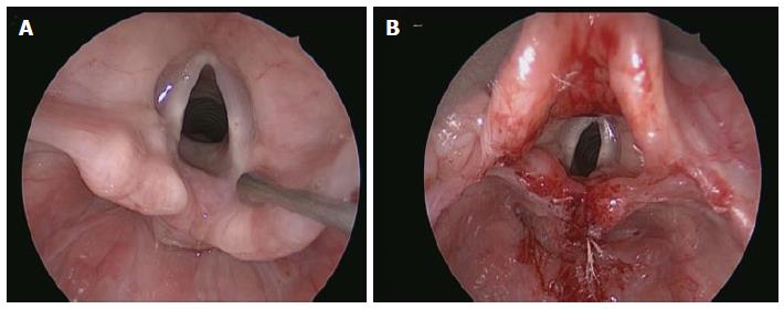
Figure 1 Endoscopic view.
A: Type 1 Laryngeal cleft; B: Endoscopic repair.
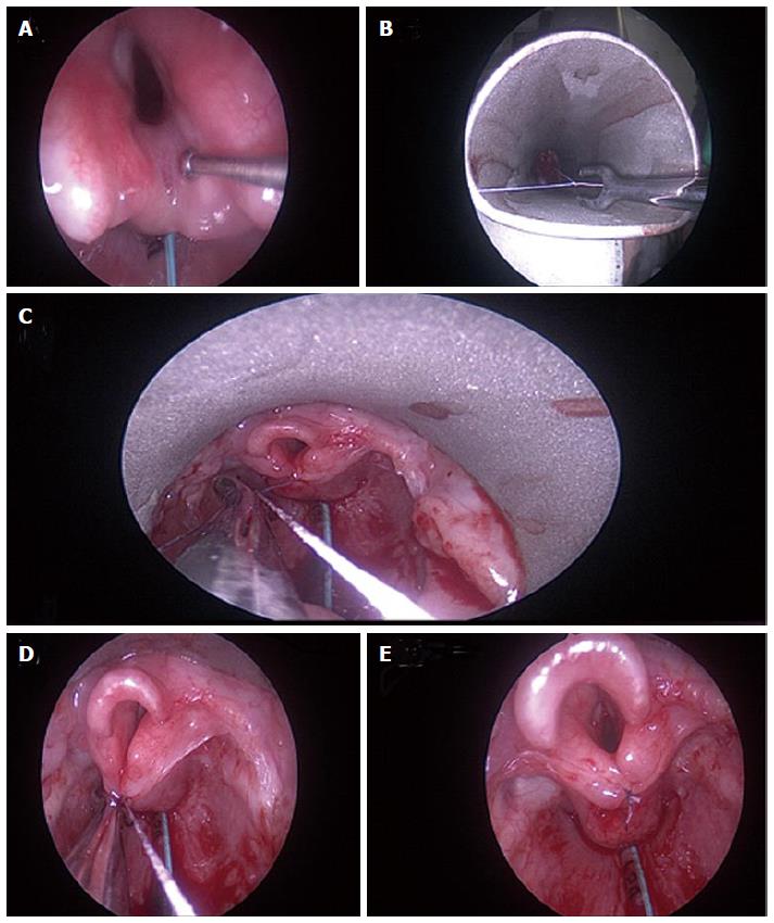
Figure 2 Novel endoscopic suture technique.
A: Type 1 Laryngeal cleft; B: Baby Benjamin suspension with Negus knot pusher and suture in situ; C, D: Squaring of knot at depth with more reliability and ease; E: Post-operative result.
- Citation: Yalamachili S, Virk JS, Bajaj Y. Diagnosis and management of laryngeal cleft: A single centre experience and a novel endoscopic technique. World J Otorhinolaryngol 2015; 5(4): 105-109
- URL: https://www.wjgnet.com/2218-6247/full/v5/i4/105.htm
- DOI: https://dx.doi.org/10.5319/wjo.v5.i4.105


