Copyright
©2013 Baishideng.
World J Otorhinolaryngol. Aug 28, 2013; 3(3): 108-113
Published online Aug 28, 2013. doi: 10.5319/wjo.v3.i3.108
Published online Aug 28, 2013. doi: 10.5319/wjo.v3.i3.108
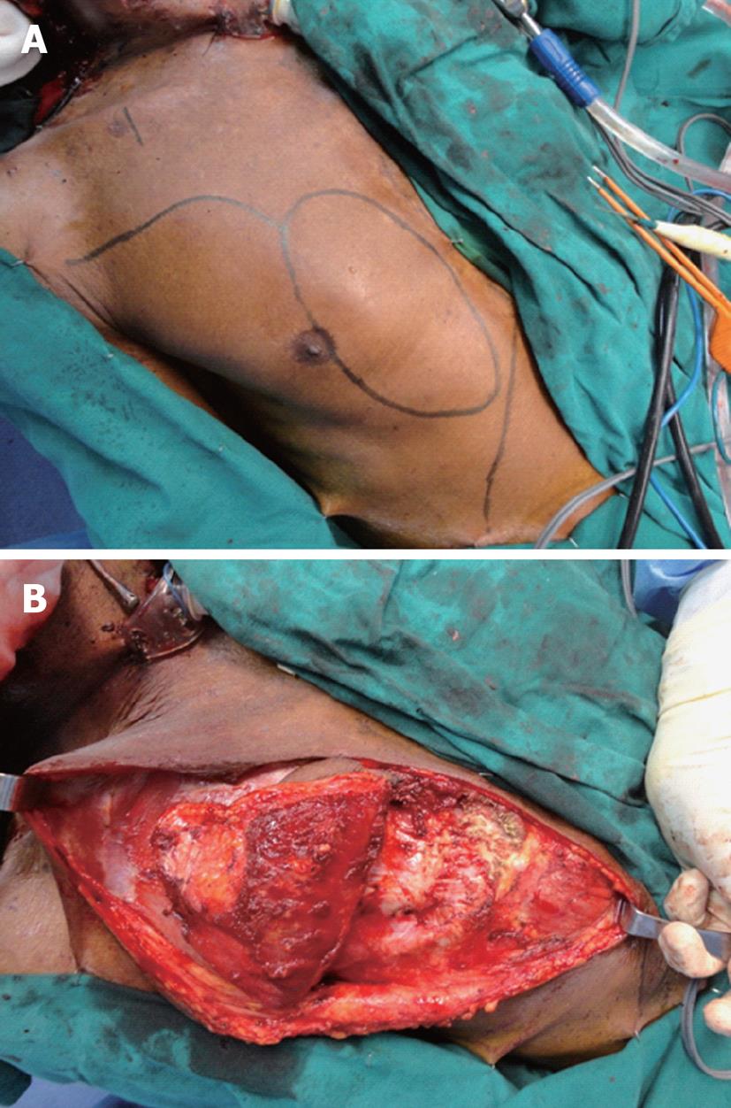
Figure 1 Flap design and elevation.
A: Marking of skin paddle with inferior line representing costal margin; B: Flap being elevated along with rectus sheath (exposed rectus muscle inferiorly).
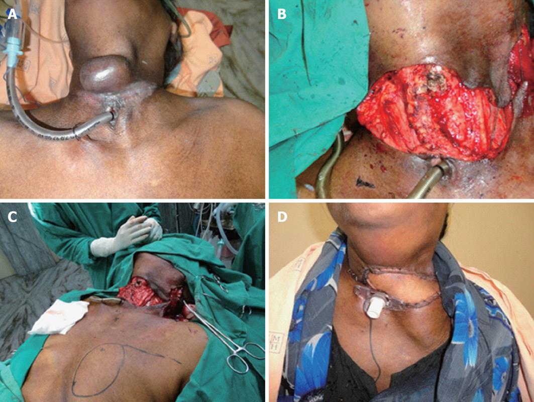
Figure 2 Reconstruction of large cutaneous defect (Patient 1).
A: Large locally invasive thyroid malignancy with skin involvement; B: Post-ablative defect; C: Marking for flap; D: Wound 1 wk after surgery.
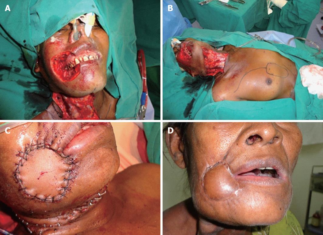
Figure 3 Reconstruction of full thickness cheek defect (Patient 3).
A: Post-ablative defect; B: Marking for skin paddle; C: Immediate post-reconstruction; D: Two months following surgery.
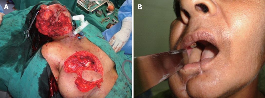
Figure 4 Reconstruction of large mucosal defect (Patient 4).
A: Elevation of flap; B: Two months following surgery.
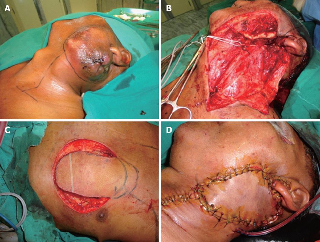
Figure 5 Reconstruction of large parotid skin defect (Patient 6).
A: Tumor parotid with skin involvement; B: Post-ablative defect; C: Skin paddle with marked line representing approximate level of inferior border of pectoralis major muscle; D: Immediately following reconstruction.
- Citation: Dhiwakar M, Nambi G. Extended pectoralis major myocutaneous flap in head and neck reconstruction. World J Otorhinolaryngol 2013; 3(3): 108-113
- URL: https://www.wjgnet.com/2218-6247/full/v3/i3/108.htm
- DOI: https://dx.doi.org/10.5319/wjo.v3.i3.108


