Copyright
©The Author(s) 2016.
World J Ophthalmol. May 12, 2016; 6(2): 10-19
Published online May 12, 2016. doi: 10.5318/wjo.v6.i2.10
Published online May 12, 2016. doi: 10.5318/wjo.v6.i2.10
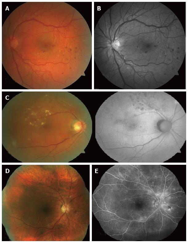
Figure 1 Retinal photographs by various funds cameras.
A: Colour fundus photo showing left eye with proliferative diabetic retinopathy; (Image taken by Zeiss Cirrus photo 800). Compare with Figure 1B; B: Red-free fundus photo (left eye with proliferative diabetic retinopathy). Better visualisation of new vessels and retinal haemorrhages as opposed to Figure 1A; C: Colour fundus photo (left) vs fundus autofluorescence (right); Retinal exudates are more visible on the colour fundus photo. Autofluorescence highlights the laser scars not readily visible on a colour fundus photo; D: Seven-field fundus photo - computer-aided mosaic (Image taken by Zeiss Cirrus 800); E: Seven-field fundus with fluorescein angiography (Image taken by Zeiss Visucam D500).
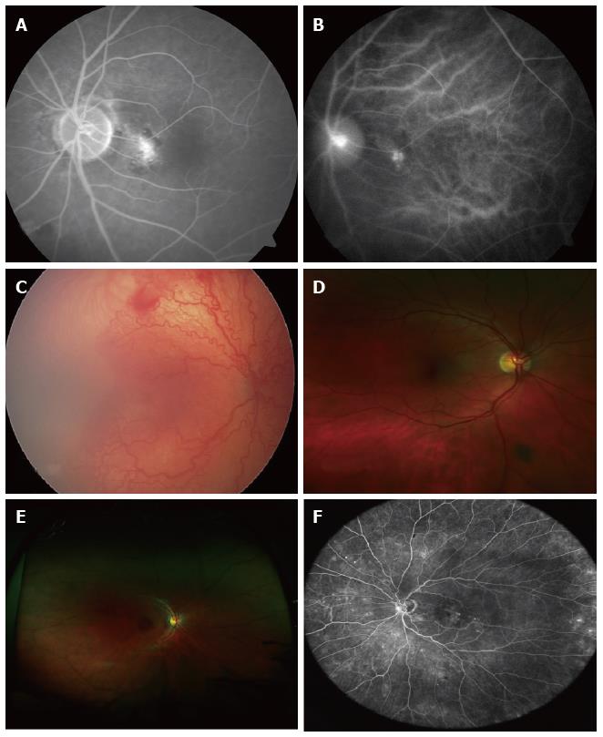
Figure 2 Advanced retinal imaging with and without contrast.
A: Fluorescein angiography. Localised hyperfluorescence; B: ICG revealing the source of leakage depicted on FFA, Figure 2A: Idiopathic polypoidal choroidal vascularisation; C: UWF fundus photo showing ROP taken with Retcam; D: Optomap ultra-widefield retinal image. Peripheral pigmented lesion not visible on standard colour fundus photo; E: Optomap ultra-widefield retinal image. Note lash artefact; F: Heidelberg HRA ultra-widefield retinal image. ICG: Indocyanine green; FFA: Fundus fluorescein angiography; UWF: Ultra-wide field; ROP: Retinopathy of prematurity.
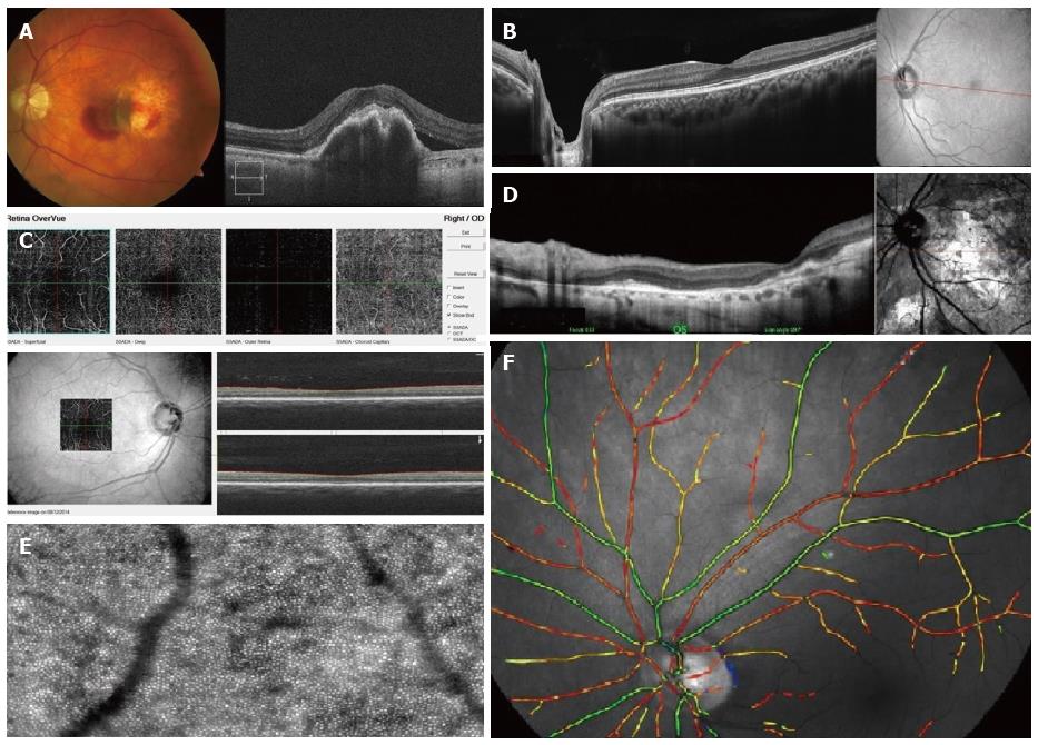
Figure 3 Retinal pictures and optical coherent tomography of various types of optical coherent tomographs showing the width and depth of image captured by each machine.
Note difference in level of detail and depth and also the accompanying retinal pictures. A: RPE rip. Colour fundus photo (left), spectral domain OCT (right). Both images taken by Zeiss Cirrus 800. This machine can obtain colour fundus photos and spectral domain OCT; B: Enhanced depth OCT showing author’s left eye (standard display includes optic disc and macula). Heidelberg OCT. Note better visualization of retinal layers; C: Non-invasive vascular imaging acquired by OptoVue OCT is spectral domain but images of the vascular tree at macula and within choroid can be seen. “Angio-OCT”; D: cSLO OCT. Good visualization of retinal layers in a patient with advanced and variable scarring; E: Adaptive optics imaging showing individual photoreceptors and microvasculature of the retina. Courtesy of imagine eyes; F: Oxymetric imaging. Courtesy of Oxymap. OCT: Optical coherent tomography; cSLO: Confocal scanning laser ophthalmoscope; RPE: Retinal pigment epithelium.
- Citation: Saeed MU, Oleszczuk JD. Advances in retinal imaging modalities: Challenges and opportunities. World J Ophthalmol 2016; 6(2): 10-19
- URL: https://www.wjgnet.com/2218-6239/full/v6/i2/10.htm
- DOI: https://dx.doi.org/10.5318/wjo.v6.i2.10


