
















©2013 Baishideng Publishing Group Co.
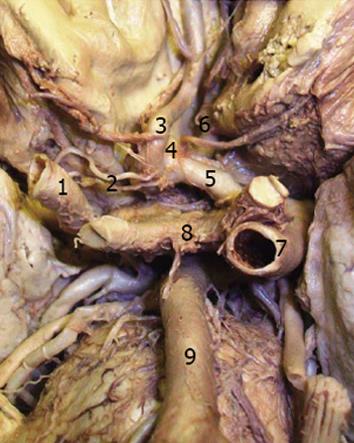
Figure 1 Duplicated hypoplastic A1 segment of the right anterior cerebral artery.
1: Right internal carotid artery (ICA); 2: Duplicated hypoplastic A1 segment of the right anterior cerebral artery (ACA); 3: A2 segment of the right ACA; 4: Anterior communicating artery (aneurysm); 5: A1 segment of the left ACA; 6: A2 segment of the left ACA; 7: Left ICA; 8: Optic chiasm; 9: Basilar artery.
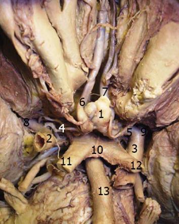
Figure 2 Duplicated hypoplastic A1 segment of the right anterior cerebral artery and anterior communicating artery aneurysm.
1: Anterior communicating artery aneurysm; 2: Right internal carotid artery (ICA); 3: Left ICA; 4: Duplicated hypoplastic A1 segment of the right anterior cerebral artery (ACA); 5: A1 segment of the left ACA; 6: A2 segment of the right ACA; 7: A2 segment of the left ACA; 8: Right middle cerebral artery (MCA); 9: Left MCA; 10: Optic chiasm; 11: Right optic nerve; 12: Left optic nerve; 13: Basilar artery.
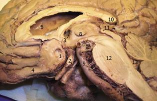
Figure 3 The depression on the internal surface of the right frontal lobe caused by anterior communicating artery aneurysm.
1: Cortical surface depression caused by aneurysm; 2: Optic chiasm; 3: Anterior commissure; 4: Gyrus rectus; 5: Right anterior cerebral artery; 6: Right lateral ventricle; 7: Right Monro foramen; 8: Massa intermedia; 9: Basilar artery; 10: Corpus callosum (splenium); 11: Pineal gland; 12: Pons (pontine nuclei); 13: Fourth ventricle.
- Citation: Mavridis IN, Anagnostopoulou S. Anterior communicating artery aneurysm associated with duplicated hypoplastic right A1 segment. World J Neurol 2013; 3(2): 10-13
- URL: https://www.wjgnet.com/2218-6212/full/v3/i2/10.htm
- DOI: https://dx.doi.org/10.5316/wjn.v3.i2.10